Expert Diagnosis, Injection Therapy and Knee Replacement for Knee Arthritis and Joint Degeneration
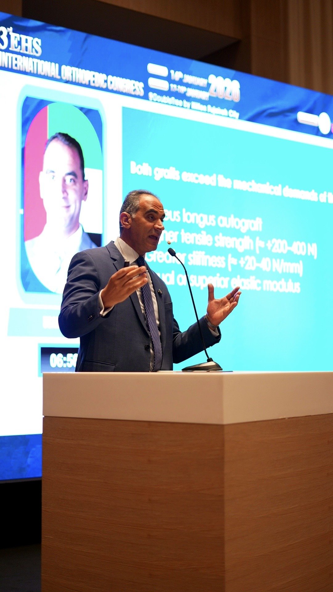
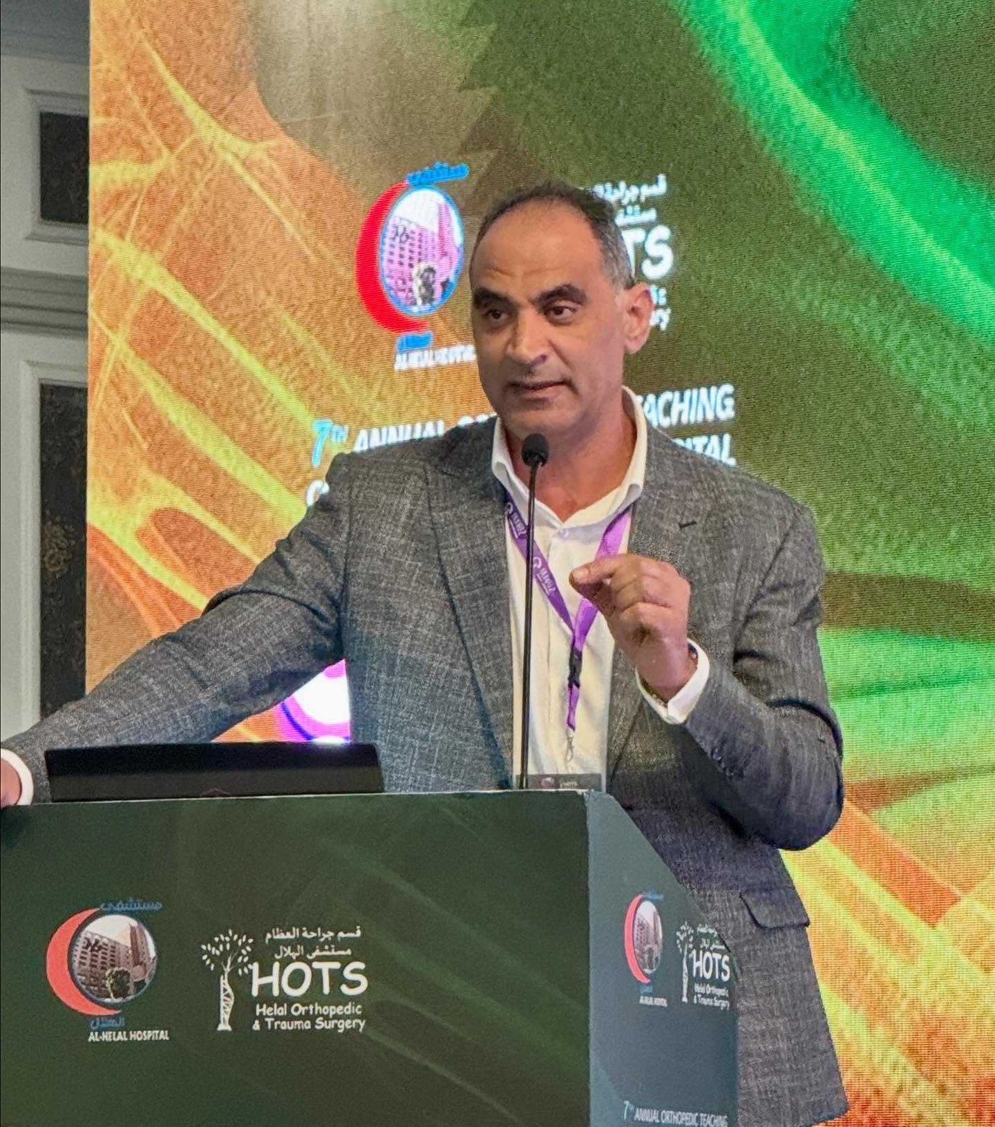
Stiffness, joint pain, and grinding sensations shouldn't limit your daily movement. Dr. Usama Saleh, a fellowship-trained orthopedic surgeon with 23 years of experience, provides personalized treatment plans tailored to your lifestyle—from targeted injection therapies to advanced knee replacement.
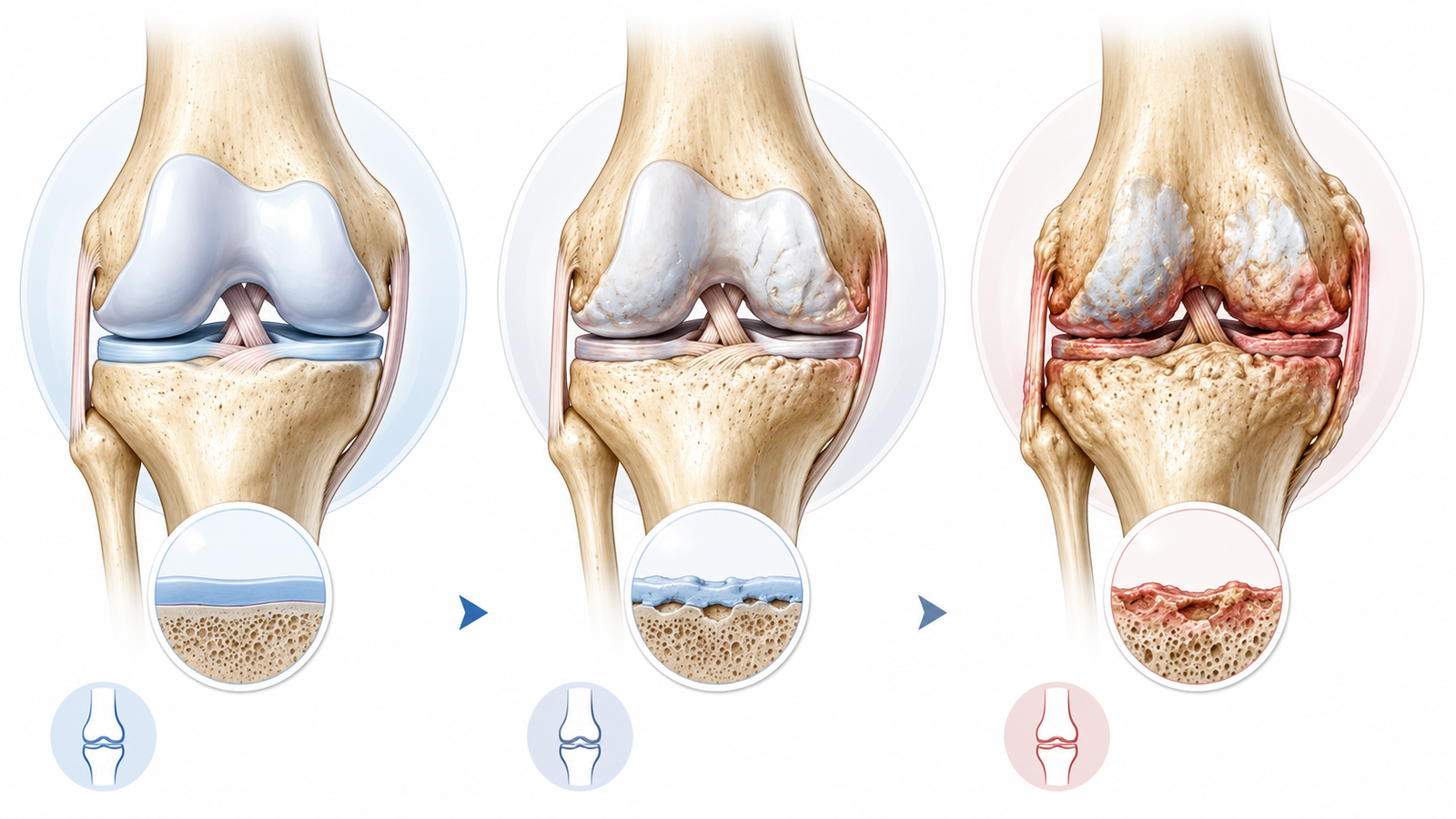
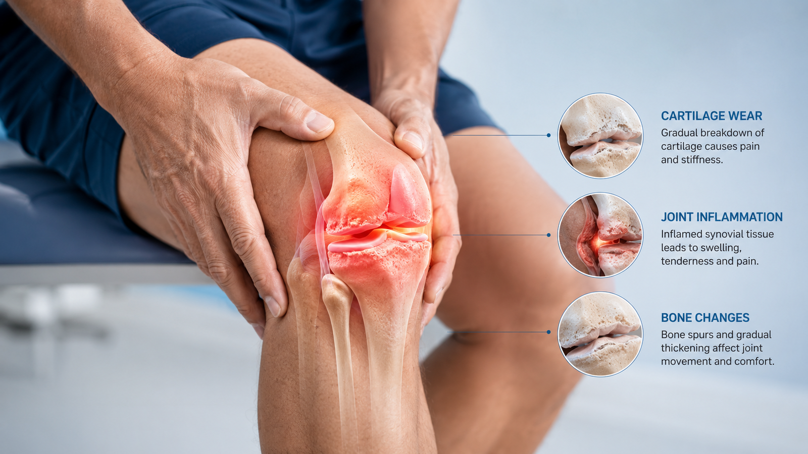
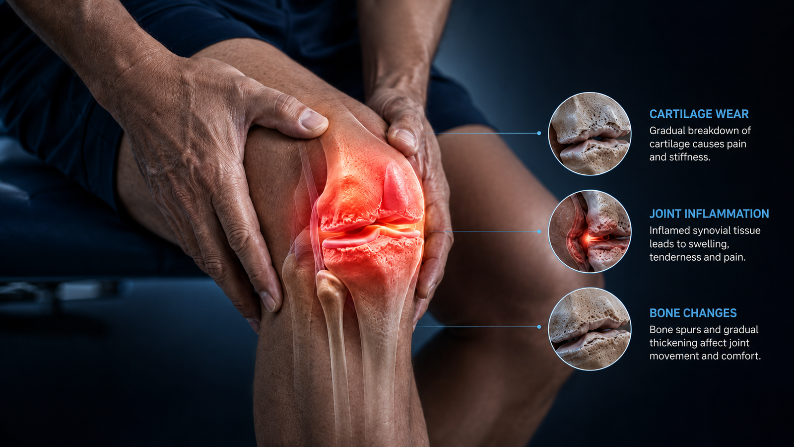
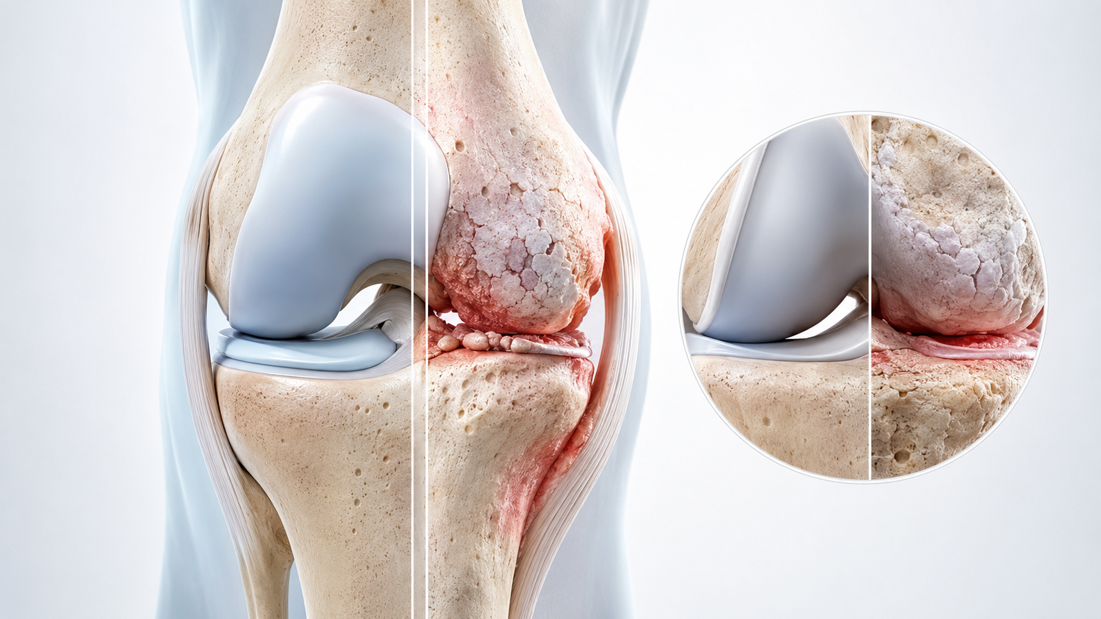
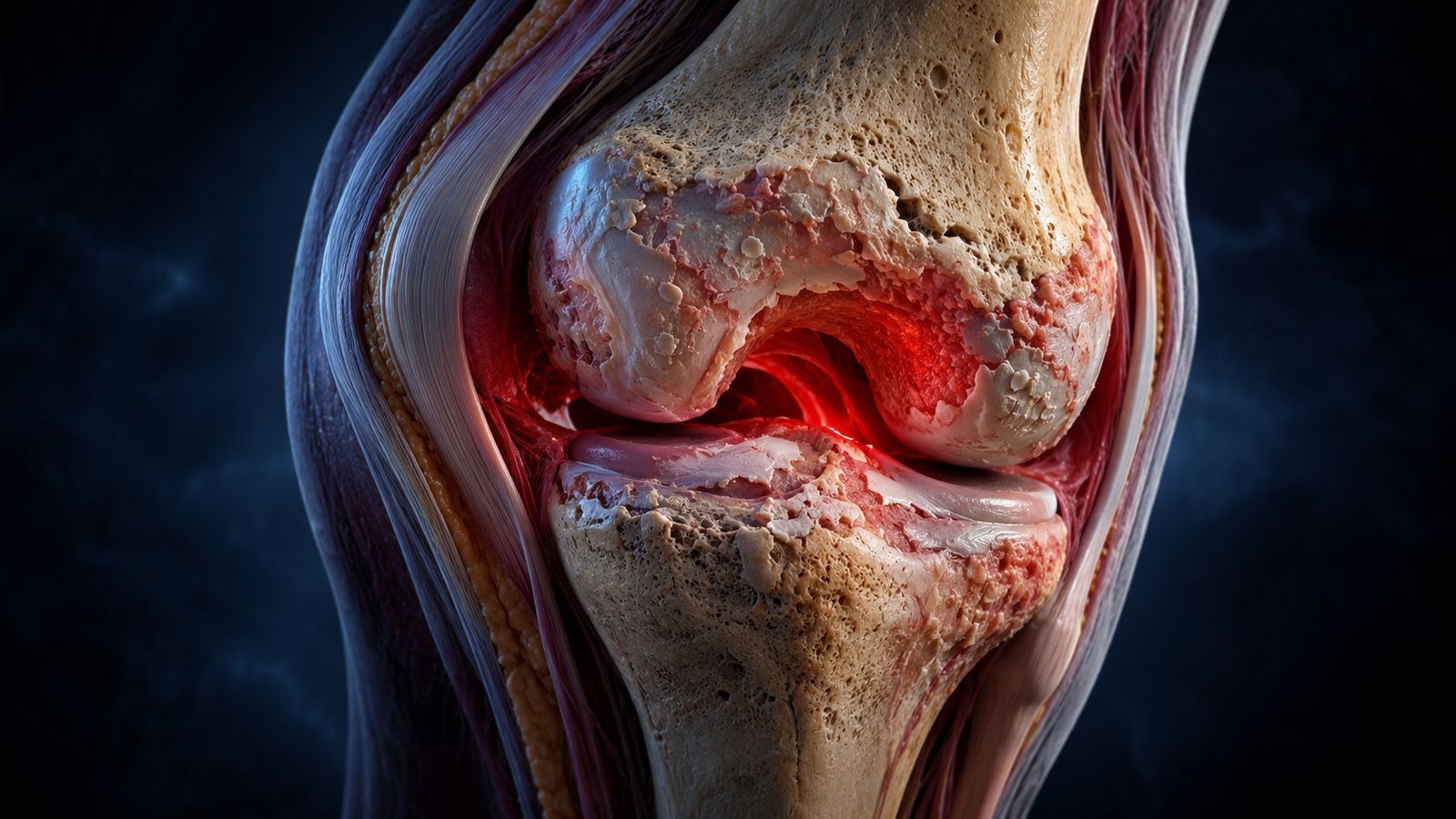
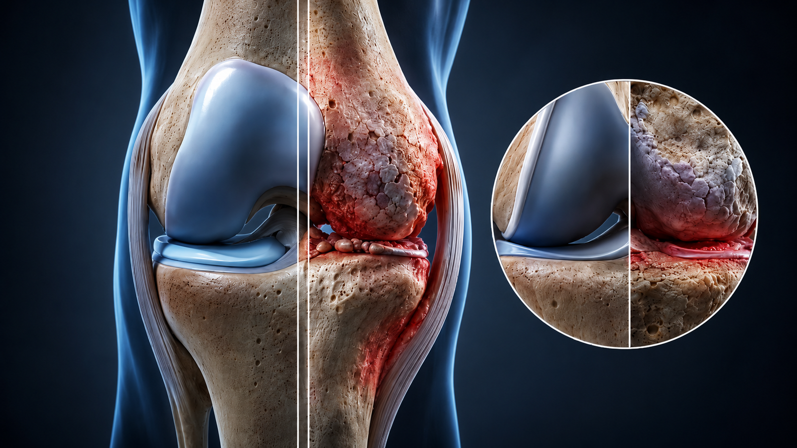
Knee osteoarthritis is a progressive degenerative joint disease caused by the gradual breakdown of articular cartilage, the smooth protective tissue covering the ends of the femur, tibia, and patella where they meet at the knee joint. As cartilage thins and erodes, the cushioning and shock-absorbing function it provides diminishes. In advanced stages, the underlying subchondral bone becomes exposed, and the surfaces of the knee joint contact each other directly, causing the characteristic bone-on-bone pain of late-stage knee osteoarthritis. Understanding knee osteoarthritis fully helps patients make informed decisions about treatment, from early physiotherapy and knee gel injections cost comparison to knowing when knee replacement surgery becomes the right step.
Osteoarthritis is distinct from rheumatoid arthritis. Rheumatoid arthritis is an autoimmune condition affecting multiple joints symmetrically. Knee osteoarthritis is a mechanical, degenerative condition driven by wear, load, age, and prior injury. Correct diagnosis determines treatment: an orthopedic specialist is the appropriate first referral for suspected knee osteoarthritis in active, working-age adults.
The knee has three compartments: the medial (inner), lateral (outer), and patellofemoral (kneecap to thigh bone). Osteoarthritis can affect one compartment (unicompartmental), two compartments (bicompartmental), or all three (tricompartmental). The medial compartment is most commonly affected first, often producing the characteristic varus (bowleg) deformity as cartilage loss progresses. Treatment selection depends significantly on which compartments are involved and to what degree.
Treatment selection is anatomy-specific. The number of affected compartments and the degree of cartilage loss help determine whether conservative care, injection therapy, partial replacement, or total knee replacement is most appropriate.
The most frequently affected compartment. Progressive cartilage loss here can produce a characteristic varus (bowleg) deformity.
Located on the outer side of the knee joint. It can be affected individually or in combination with other compartments.
Positioned between the kneecap and thigh bone. Wear here typically causes severe pain during stairs, squatting, or standing up.
Knee osteoarthritis is graded using the Kellgren-Lawrence (K-L) classification on weight-bearing X-rays. Understanding your stage guides every treatment decision, from conservative management to injection therapy to knee replacement timing:
Grade 01
Minimal joint space narrowing on X-ray. Minor osteophyte formation with microscopic cartilage softening.
Often mild or absent. Occasional morning stiffness or mild aching after strenuous exercise.
Grade 02
Noticeable osteophyte formation and definite joint space narrowing. Cartilage begins to thin.
Intermittent pain during movement, morning stiffness under 30 minutes, and joint crepitus.
Grade 03
Significant joint space narrowing, multiple osteophytes, and obvious areas of cartilage erosion.
Frequent pain with walking or stairs, recurrent swelling episodes, and reduced mobility range.
Grade 04
Bone-on-bone contact, severe space loss, large osteophytes, and marked limb malalignment.
Constant, severe pain resting or waking at night, with severe daily movement limitation.
Pain worse after activity and better with rest: the classic early OA pattern, settling after sitting or lying down.
Morning stiffness lasting under 30 minutes: a key distinguishing feature of OA compared to rheumatoid arthritis.
Crepitus: a grinding or grating sensation during knee movement from roughened cartilage surfaces.
Intermittent mild swelling after prolonged activity, settling with rest and ice applications.
Constant pain not relieved by position change, disturbing sleep and indicating advanced cartilage loss.
Joint effusion from chronic synovial inflammation, causing warmth and often requiring fluid aspiration.
Development of varus (bowleg) or valgus (knock-knee) alignment as compartmental cartilage erodes.
Giving way sensation and severe limitation in walking distance or fully extending the knee joint.
Two types of knee osteoarthritis: Primary OA develops without a specific cause, driven by age-related cartilage degeneration and accumulated mechanical wear. Secondary OA develops after a specific joint insult, with post-traumatic OA after ACL tear, meniscal loss, or fracture being particularly common in Dubai's active population.
Primary OA develops without a specific cause, driven by age-related cartilage degeneration and accumulated mechanical wear.
Secondary OA develops after a specific joint insult, with post-traumatic OA after ACL tear, meniscal loss, or fracture being particularly common in Dubai's active population.
Age over 45: cartilage repair capacity declines with age, making cumulative wear progressively more difficult to reverse.
Previous knee injury: prior ACL tear, meniscal loss after meniscectomy, or tibial plateau fracture dramatically accelerates OA. Post-meniscectomy knees develop OA at 10-15 years at significantly higher rates.
Obesity: Every kilogram of body weight creates approximately 4 kilograms of force across the knee joint during walking. A 10-kilogram excess translates to 40 kilograms of additional knee joint loading per step.
Sedentary or occupational overuse: prolonged sitting weakens the quadriceps, reducing dynamic knee protection. Occupational kneeling and squatting accelerates medial compartment wear.
Female sex: women develop knee OA at higher rates than men, particularly after age 50 due to hormonal and anatomical differences.
Limb malalignment: varus or valgus alignment concentrates load disproportionately in one compartment, accelerating unilateral cartilage wear.
Diagnosis combines clinical examination with weight-bearing imaging. Non-weight-bearing X-rays significantly underestimate joint space narrowing and should not be used for OA staging.
Non-weight-bearing X-rays significantly underestimate joint space narrowing and should not be used for OA staging.
The selected imaging method depends on disease stage, symptoms, age, associated injuries, and the intended treatment.
Gait analysis for varus or valgus thrust. Standing limb alignment. Joint line palpation for tenderness. ROM assessment. Crepitus. Ligament stability testing to exclude coexisting instability. Kellgren-Lawrence staging from X-ray.
Gold standard for OA diagnosis and staging. Bilateral AP (standing), lateral, and skyline (patellofemoral) views. Joint space narrowing, osteophytes, subchondral sclerosis, and cysts are assessed using the K-L grading scale (Grade 1-4).
Bilateral Standing Joint-space assessment under body load
Lateral View Bone profile and alignment assessment
Skyline View Patellofemoral compartment assessment
Not routinely required for diagnosis but valuable for assessing cartilage quality in younger patients, identifying coexisting meniscal tears (present in up to 60% of OA knees), subchondral bone edema, and surgical planning for partial vs. total knee replacement.
Coexisting meniscal tears Present in up to 60% of OA knees
Used in clinic to assess joint effusion, confirm injection targets, and guide gel injection procedures. Ultrasound-guided injections are significantly more accurate than landmark-guided injections, particularly in obese patients.
Joint Effusion Assesses fluid accumulation
Injection Target Confirms exact treatment location
Gel Injection Guidance Real-time needle positioning
Ultrasound-guided injections are significantly more accurate than landmark-guided injections, particularly in obese patients.
Examine Clinical Findings
Stage Weight-Bearing X-ray
Investigate MRI When Needed
Guide Ultrasound Procedure
Plan Personalized Treatment
Treatment is stage-specific, age-specific, and activity-specific. Dr. Usama presents every option transparently, starting with the most conservative effective approach and proceeding to surgical intervention only when conservative management no longer provides adequate function.
Physiotherapy, weight reduction, and activity modification for joint load reduction.
Gel viscosupplementation, Corticosteroid, or PRP biological therapy.
Partial or Total Knee Replacement for definitive, long-term functional recovery.
Targeted quadriceps strengthening reduces pain and improves knee function in 60–80% of early-to-moderate OA cases.
60–80% Function ImprovementA 5–10% body weight reduction significantly reduces mechanical pressure across the joint and slows disease progression.
4kg Force Saved per 1kg LostTransitioning to low-impact exercise such as swimming, cycling, and water walking protects the cartilage while building strength.
Injecting hyaluronic acid restores natural synovial fluid lubrication and shock absorption. Typically provides 6–12 months of relief and is covered by major UAE insurance providers.
Rapid anti-inflammatory relief for severe flare-ups. Provides 6–12 weeks of comfort, acting as an effective bridge to active rehab.
Concentrated growth factors from your own blood stimulate natural tissue healing and reduce inflammation in early-to-moderate stages.
The gold standard for end-stage bone-on-bone osteoarthritis. Resurfaces damaged joint surfaces with advanced implants, restoring pain-free mobility with a 90–95% satisfaction rate at 10 years.
Ideal for isolated single-compartment damage. Preserves healthy ligaments and cartilage, offering a smaller procedure with faster post-operative recovery.
We prioritize joint preservation first through structured non-surgical pathways, escalating to replacement only when necessary for your mobility and quality of life.
The most important reason to seek early specialist assessment is not to rush into treatment. It is to get an accurate stage diagnosis that determines which treatments are appropriate now and which may be needed later.
If your knee suddenly becomes severely painful, swollen, and warm to the touch without prior trauma. This requires urgent evaluation to rule out septic arthritis, acute gout, or a severe flare-up.
Inability to walk or stand following a fall or minor impact. Arthritic bone can suffer micro-fractures under lighter trauma than healthy joint bone structure.
Continuous or activity-related pain that hasn't improved with rest, limiting daily walking distance, stairs, or recreation.
Relying on over-the-counter painkillers or anti-inflammatories for over a month to manage basic daily activities.
Stiffness taking more than half an hour to ease every morning, or joint aching waking you up from rest/sleep.
Seeking early assessment protects your access to effective non-surgical treatments—like joint preservation physiotherapy and viscosupplementation injections—before options narrow.
CAUTION: Do NOT exercise if your knee is acutely swollen or significantly more painful than usual. Exercise during an acute flare increases inflammation and can worsen cartilage damage. Wait until the acute episode resolves. After injections: rest the injected knee for 24-48 hours after corticosteroid or hyaluronic acid injections and 48-72 hours after PRP. If you have been advised that knee replacement is needed, consult Dr. Usama before starting a new exercise program.
Quad sets: sit or lie with legs straight. Tighten the quadriceps by pressing the back of the knee toward the floor, hold for 5 seconds, and release. 3 sets of 15 daily. The single most important exercise for knee OA, directly reducing joint load by strengthening the primary knee stabilizer.
Straight leg raises: lie on your back, one knee bent, the affected leg straight. Tighten the quad and raise the straight leg to knee height. Hold for 3 seconds, lower slowly. 3 sets of 15. Strengthens quads without compressive joint loading.
Short arc quads: lie with a rolled towel under the knee, creating 30 degrees of flexion. Straighten the leg fully against gravity, hold for 3 seconds, and lower slowly. 3 sets of 15. Targets terminal quadriceps extension critical for normal walking mechanics.
Cycling (stationary or outdoors): the single best cardiovascular exercise for knee OA. Low-impact, joint-friendly, and directly strengthens the quadriceps. Adjust saddle height to avoid full knee flexion. 20-30 minutes, 3-4 times per week.
Water walking or hydrotherapy: walking in chest-deep water reduces knee joint load by approximately 75% while maintaining cardiovascular and strengthening benefits. Ideal for moderate-to-severe OA patients unable to tolerate land-based exercise.
Running and jogging: high-impact loading accelerates cartilage wear in moderate-to-severe OA. Replace with cycling, swimming, or water walking.
Deep squats and lunges: compressive loading on the patellofemoral joint is highest in deep flexion, particularly damaging in OA with patellofemoral involvement.
Leg press with a heavy load through the full range of motion: light leg press at 0-45 degrees is acceptable. Heavy loading is contraindicated.
High-impact sports (football, basketball, tennis, and padel) in moderate-to-severe OA. Low-impact alternatives such as golf, cycling, and swimming are appropriate for most OA patients.
Assessment and planning (15 minutes): Dr. Usama confirms the target knee and injection site using clinical examination and reviews the most recent X-ray. The injection approach is selected for optimal joint access, most commonly the superolateral or lateral mid-patellar approach.
Patient positioning (2-3 minutes): patient lies on the treatment table with the affected knee relaxed in slight flexion. The area is cleaned with antiseptic solution. A local anesthetic spray or subcutaneous lidocaine injection numbs the entry site.
Ultrasound setup (2-3 minutes): a real-time ultrasound transducer is positioned to visualize the suprapatellar pouch above the kneecap. Ultrasound guidance ensures the needle enters the joint space accurately, significantly improving precision versus landmark-guided injection, particularly in obese patients.
Real-time visualization improves needle accuracy compared with landmark guidance.
Aspiration if effusion is present (2-5 minutes): if the knee contains excess fluid (joint effusion), Dr. Usama aspirates it through the same needle before injecting the gel. Removing excess fluid improves both the comfort and efficacy of viscosupplementation.
Joint finding Excess Fluid Present
Procedure Aspiration
Result Better Comfort
Treatment benefit Improved Efficacy
Gel injection (1-2 minutes): the hyaluronic acid gel is injected into the joint space under real-time ultrasound visualization, coating the cartilage surfaces. The needle is removed and a small adhesive dressing applied.
Hyaluronic acid coats the cartilage surfaces and supplements natural joint lubrication.
Post-injection protocol (5 minutes): brief rest in clinic. Avoid high-impact activities for 24-48 hours. Ice for 15 minutes if mild soreness develops. Full benefit from hyaluronic acid typically develops over 4-6 weeks as the gel integrates with the joint environment.
30-45 minutes total at Medcare MOSH including consultation and post-injection rest
Local only: no general anaesthesia, no overnight stay
Same-day return to desk work
After 24-48 hours rest
Every 6-12 months depending on response and product used
AED 1,200-3,000 per session: most UAE insurance plans cover for confirmed OA
Achieved in Stage 1–2 OA with structured physiotherapy. Benefits develop over 8–12 weeks of consistent adherence.
Meaningful pain reduction in moderate OA. Benefits peak at 8–12 weeks, with a duration of 6–12 months. Re-injectable every 6 months. Among the most cost-effective non-surgical OA interventions available.
Measured at 6 months for Stage 1–3 OA with a single session every 6–12 months. Shows a greater biological effect in younger patients with early disease.
One of the highest satisfaction rates of any elective surgical procedure when appropriately timed.
All injection procedures at Medcare MOSH are performed under ultrasound guidance with strict sterile technique to minimize procedural risks. Dr. Usama believes complete transparency about potential risks serves patients far better than reassurance that minimizes genuine considerations.
Two decades of subspecialty, fellowship-level expertise from diagnosis to advanced non-surgical injection therapy and complex knee replacement surgery under one roof at Medcare MOSH.
Fellowship-Trained Knee Specialist at Medcare MOSH
Advanced fellowship training in orthopedic surgery at the University of Toronto providing subspecialty expertise in the full spectrum of knee OA management: conservative protocols, injection therapy, and complex knee replacement. Fellowship-trained surgeons demonstrate consistently better patient outcomes in published literature.
Dr. Usama never recommends knee replacement before exhausting appropriate conservative and injection therapy options. Patients at Stage 2-3 OA are managed with structured physiotherapy and repeat injection cycles for as long as outcomes remain adequate, delaying surgery by years in appropriately selected patients.
From same-consultation X-ray and weight-bearing imaging to ultrasound-guided gel and PRP injections, structured physiotherapy, and knee replacement surgery: all at Medcare Orthopaedics and Spine Hospital (MOSH), Sheikh Zayed Road, Dubai. No fragmented care, no referral delays.
Two decades managing knee osteoarthritis in Dubai's diverse, active population, from younger post-traumatic OA patients in their 30s and 40s to older adults with advanced tricompartmental disease. Hundreds of knee replacements and thousands of injection procedures.
Knee OA treatment decisions, particularly the question of when knee replacement is truly necessary, are among the most important a patient makes. Dr. Usama presents staging information clearly using X-ray evidence, explains every option with honest expected outcomes, and aligns the plan with each patient's specific goals.
Active AO Trauma Faculty member, published peer-reviewed manuscripts, book chapter author, international conference presenter. Your knee OA management is informed by the most current evidence-based practices globally.


Knee osteoarthritis is a progressive degenerative joint disease caused by the gradual breakdown of articular cartilage in the knee joint. As cartilage thins and eventually wears through, the bones begin to contact each other directly, causing pain, stiffness, swelling, and loss of function. It is the most common joint condition in adults over 45 and the leading cause of knee pain in Dubai's aging active population. OA is mechanical and degenerative, distinct from rheumatoid arthritis, which is autoimmune. Treatment is managed by an orthopedic surgeon.
Yes. The majority of knee OA patients, particularly those in Stages 1-3, can be managed effectively without surgery for years. First-line treatments include structured physiotherapy with quadriceps strengthening (60-80% adequate symptom control in early-moderate OA), weight management, and activity modification. When conservative management is insufficient, injection therapy options include hyaluronic acid gel injections (viscosupplementation), corticosteroid injections, and PRP. Knee replacement is reserved for Stage 4 patients where conservative management has failed. Dr. Usama's conservative-first philosophy means surgery is recommended only when it clearly offers better outcomes than non-surgical management for a specific patient's stage and demands.
Knee OA is staged using the Kellgren-Lawrence (K-L) grading scale on weight-bearing X-rays: Stage 1 (Minor): minimal cartilage changes, mild or no symptoms. Stage 2 (Mild): early osteophytes, some cartilage thinning, and intermittent pain with activity. Stage 3 (Moderate): significant joint space narrowing, frequent pain, and activity limitation. Stage 4 (Severe): severe joint space narrowing, often bone-on-bone contact, constant pain, and significant functional limitation. Treatment is stage-specific: Stages 1-2 are managed conservatively, Stages 2-3 benefit from injection therapy and physiotherapy, and Stage 4 may require knee replacement when conservative management has been fully tried.
Dr. Usama Saleh at Medcare Orthopaedics and Spine Hospital (MOSH), Sheikh Zayed Road, Dubai, is a fellowship-trained orthopedic surgeon with 23 years of knee OA experience. His expertise spans the full spectrum of knee OA management: evidence-based conservative protocols, ultrasound-guided injection therapy (hyaluronic acid, PRP, and corticosteroid), and knee replacement surgery for end-stage disease. His conservative-first philosophy means patients receive an honest assessment of whether and when surgery is truly necessary rather than being referred prematurely. If you are searching for a knee pain doctor near me in Dubai, Dr. Usama's team can confirm same-day appointment availability.
Knee gel injection cost is one of the most common questions patients ask when exploring non-surgical OA management in Dubai.
Knee gel injection costs in Dubai typically range from AED 1,200 to AED 3,000 per session depending on the specific hyaluronic acid product used (single-injection vs. multi-injection protocols) and the clinic. Single-injection formulations are more convenient but cost more per session. Multi-injection protocols have lower cost per injection but higher total session cost. Most major UAE insurance plans, including Daman, DHA, Cigna, AXA, Bupa, MetLife, GlobeMed, NAS, and NextCare, cover viscosupplementation for confirmed knee osteoarthritis. Dr. Usama's team at Medcare MOSH verifies your insurance coverage before scheduling and provides transparent cost estimates upfront.
Total knee replacement in Dubai typically costs AED 60,000-100,000, including surgeon fees, implant, anesthesia, and hospital stay. Partial (unicompartmental) knee replacement: AED 50,000-80,000. Both procedures are covered by most comprehensive UAE insurance plans for patients with confirmed end-stage osteoarthritis and documented failure of conservative management. Prior insurance authorization is obtained by Dr. Usama's team before scheduling. Transparent itemized cost estimates are provided during consultation for all knee replacement procedures at Medcare MOSH Dubai.
Untreated knee osteoarthritis is progressive. Without management, cartilage loss continues, joint space narrows, and bone-on-bone contact develops. Secondary consequences include joint deformity (varus or valgus), quadriceps muscle atrophy from offloading the painful knee, chronic pain sensitization, and progressive loss of walking ability. Patients presenting at Stage 4 after years of untreated Stage 2-3 OA often face more complex surgery with worse functional outcomes than patients who managed their OA proactively. Earlier specialist assessment consistently produces better long-term outcomes.
Some supplements have moderate evidence supporting symptom improvement in knee OA. Glucosamine and chondroitin sulfate: meta-analyses show modest pain reduction in a subset of OA patients, particularly moderate OA. Low risk. Omega-3 fatty acids (fish oil): anti-inflammatory properties with supporting evidence for joint pain reduction. Collagen peptides: emerging evidence for early OA symptom management. Vitamin D: deficiency is associated with OA progression; supplementation is reasonable for patients with confirmed deficiency, which is common in Dubai due to sun avoidance and office lifestyles. No supplement can reverse cartilage damage or replace appropriate orthopedic management, but as adjuncts to physiotherapy and injection therapy, several have a reasonable evidence base.
Try searching for injections, surgery, cost, stages, or supplements.
Explore the complete knee diagnosis and treatment pathway in Dubai.
Hyaluronic acid, corticosteroid, and image-guided injection options.
Definitive surgical care for advanced knee osteoarthritis.
Keyhole treatment for selected meniscal and intra-articular conditions.
Platelet-rich plasma and biological treatment for selected patients.
Structured strength, mobility, and return-to-activity rehabilitation.
Whether you have just noticed the first signs of knee stiffness or have been managing knee arthritis for years without a specialist plan, Dr. Usama Saleh at Medcare MOSH provides the accurate staging and honest treatment guidance that makes the difference between managed OA and progressive joint deterioration.
Weight-bearing X-ray review and Kellgren-Lawrence staging at your first appointment
Clear explanation of your OA stage and exactly what it means for your treatment options
Conservative, injection, and surgical options presented transparently with realistic timelines
Personalised physiotherapy and exercise plan aligned with your activity level and goals
Insurance verification and transparent knee gel injections cost information provided before any procedure, including whether your UAE plan covers viscosupplementation
Second opinion welcome: honest assessment of whether knee replacement is truly necessary at your current stage
World-Class Trauma and Sports Injury Surgery — Tailored to You.
Powered by ICONCLY Medical.