Frozen Shoulder Treatment Dubai: Restore Movement & End Pain

Progressive shoulder stiffness making it impossible to reach behind your back, severe night pain disrupting sleep, and loss of simple daily activities like brushing hair or getting dressed—these are the frustrating hallmarks of frozen shoulder, medically termed adhesive capsulitis. This condition causes the shoulder joint capsule to thicken and contract, creating profound stiffness and pain that typically progresses through three distinct frozen shoulder stages over 12-24 months. Dr. Usama Saleh, Dubai’s fellowship trained shoulder specialist with 23+ years of experience, provides expert frozen shoulder treatment focused on accelerating recovery through targeted physical therapy, frozen shoulder exercises, corticosteroid injections, and gentle manipulation.

Understanding Frozen Shoulder
Capsular Pathologies

Understanding
Frozen Shoulder

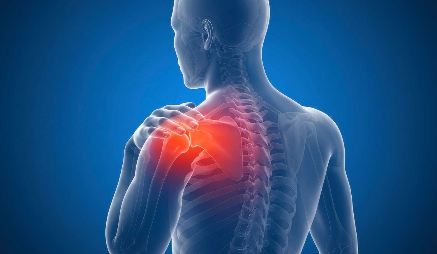
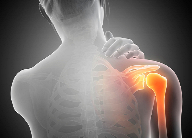
Frozen shoulder, clinically termed adhesive capsulitis, is a highly restrictive pathology targeting the connective tissue envelope of the joint.

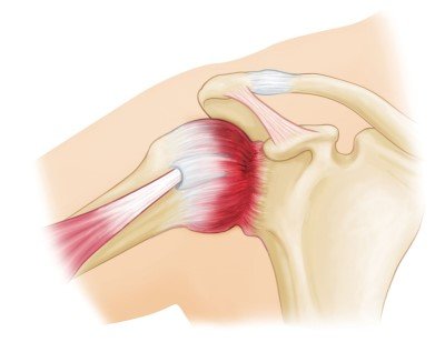
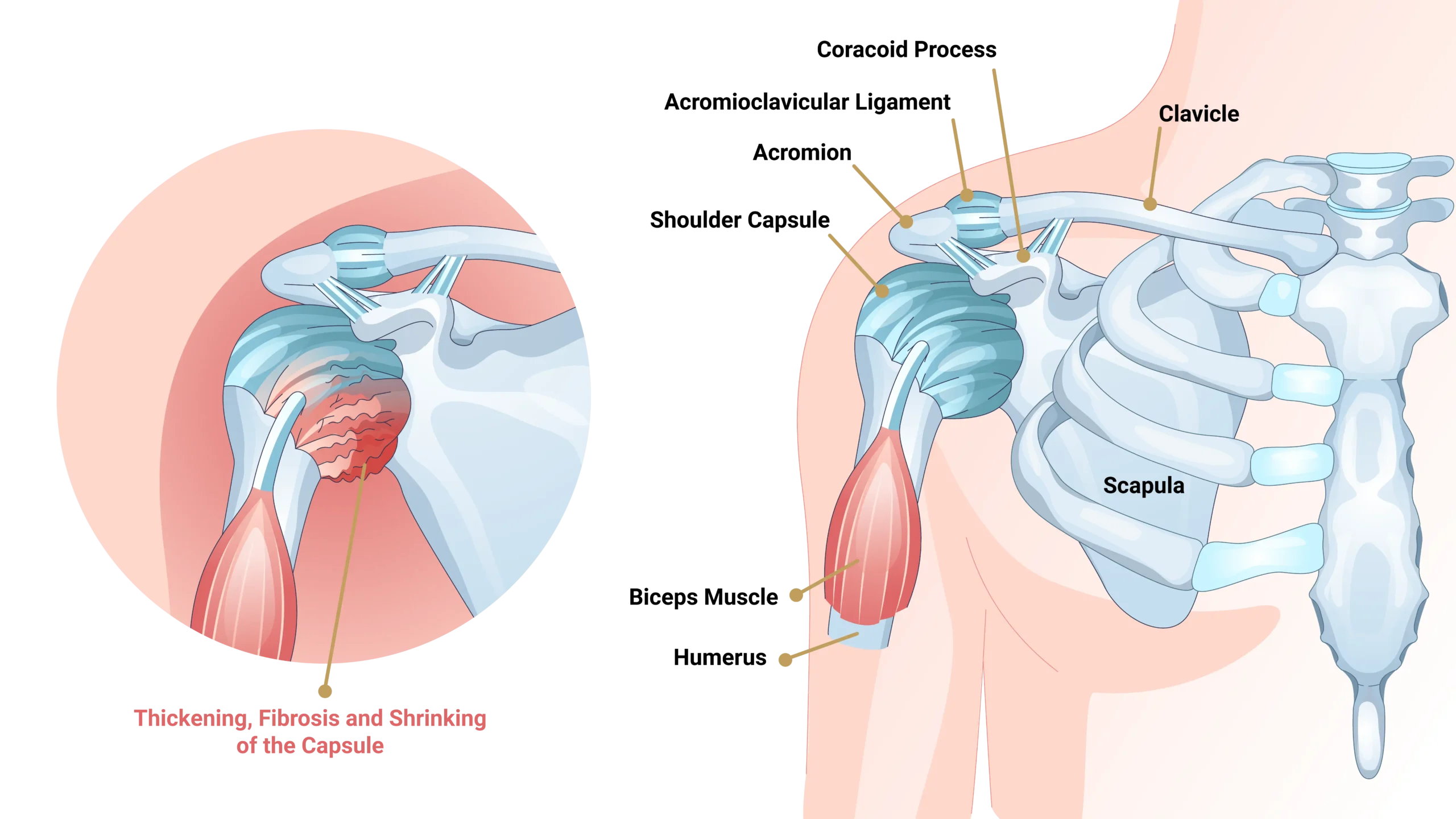
The condition occurs when the shoulder joint capsule becomes profoundly inflamed, thickened, and contracted. This global capsular tightening severely restricts both active movement (you moving your arm) and passive movement (someone else moving your arm).

The Unique Diagnostic Indicator:

Unlike most subacromial or rotator cuff conditions where passive motion remains intact, frozen shoulder uniquely limits movement in all operational planes with a distinct, characteristic ‘hard stop’ mechanical block upon clinical testing.
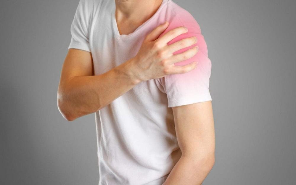
Symptom Tracking

Frozen Shoulder Symptoms

Recognizing clinical indicators early optimizes targeted care strategies. Frozen shoulder presents with a distinct mechanical progression through chronological phases.

Primary Symptoms Scan

Global ROM Restriction

Gradual, severe loss of range of motion in all directions, marked by a characteristic ‘hard stop’ when attempting movement.

Nocturnal Pain Spikes

Frozen shoulder pain becomes exceptionally severe at night, profoundly disrupting sleep cycles when lying on the affected side.

Functional Limitations

Severe mechanical difficulty executing everyday tasks like brushing hair, reaching a back pocket, or fastening garments.

Chronological Stage Progression

Stage 1: Freezing

Duration: 2-9 Months. Progressive pain worsening over weeks with active movement and at rest. Vertical limits restrict reaching overhead or behind the back tissue fields.

Stage 2: Frozen

Duration: 4-12 Months. Resting pain decreases significantly but profound global stiffness remains locked. Joint external rotation metrics are typically most affected.

%

Stage 3: Thawing & Recovery

Gradual spontaneous improvement in biological motion tracking over a 6-24 month window. Pain metrics become minimal as functional capacity slowly restores.

Clinical Recovery Outlook: Total healing time left untreated spans 18-24 months, with 10-15% of cases retaining permanent residual stiffness. Specialized targeted treatment accelerates this recovery trajectory significantly.

Diagnostic Profiling

Diagnosis Of
Frozen Shoulder

Frozen shoulder diagnosis is primarily clinical. Dr. Usama maps the characteristic restriction patterns to isolate capsular pathology from other joint conditions accurately.

Global Expertise. Local Heart.
MD PhD MRCS (UK) Fellowship-Trained (Canada)

Board certified in orthopedics surgery

Clinical Examination

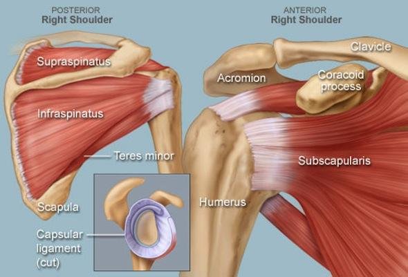
Testing reveals severe range of motion restriction in all directions, notably forward flexion, abduction, and external rotation (the most limited plane). A key differentiator is that both active and passive ROM are limited, tracking a strict 'capsular pattern' with a smooth, solid 'hard stop' at end range, completely free of crepitus or grinding.

Imaging Studies

X-rays & MRI: X-rays typically return normal parameters, serving primarily to rule out joint arthritis or calcific tendinitis. MRI can visualize a thickened joint capsule and decreased joint volume metrics but is generally not required for definitive confirmation.
Ultrasound Tracking: May show explicit coracohumeral ligament thickening. Imaging is deployed mainly as a protective check to rule out secondary rotator cuff tears if the diagnosis remains unclear.

Clinical Case 1
Clinical Case 2
Clinical Case 3
Clinical Case 4
Clinical Case 5
Clinical Case 1
Clinical Case 2
Clinical Case 3
Clinical Case 4
Clinical Case 5
Clinical Case 6
Clinical Case 7
Clinical Case 8
Clinical Case 9
Clinical Case 10
Clinical Case 6
Clinical Case 7
Clinical Case 8
.
Clinical Case 9
Clinical Case 10
Clinical Stratification

Frozen Shoulder Treatment Options

How to treat frozen shoulder focuses dynamically on reducing inflammation, maintaining joint range of motion, and accelerating the natural recovery process. Adhesive capsulitis treatment is primarily conservative, with surgery rarely required.

Protocol Phase 01

Stage 1: Freezing (Painful Phase)

Primary Treatment Target:

Minimize acute pain progression, protect baseline joint mobility, and aggressively prevent excessive capsular tightening.

Pain Control Interventions

Utilization of targeted NSAIDs combined with corticosteroid subacromial/intra-articular injections. Injections operate as an exceptionally effective measure for acute pain control and may slow the functional progression of capsular thickening.

Gentle ROM Rehabilitation

Structured daily exercises strictly avoiding any forced painful ranges. Physical therapy tracking focuses heavily on maintaining tissue mobility while adapting precise sleep positioning parameters to reduce severe night pain disruptions.

Phase Clinical Focus: Pain Relief Dominant
Protocol Phase 02

Stages 2-3: Frozen & Thawing

Primary Treatment Target:

Shatter rigid structural capsular adhesions, restore comprehensive range of motion, and accelerate the natural thawing timeline.

Aggressive Mobilization Matrix

High-compliance frozen shoulder physical therapy integrating dedicated, targeted capsular stretching. Execution requires specialized frozen shoulder exercises performed daily with perfect mechanical technique. Corticosteroid injections may be introduced to manage residual tracking pain.

Advanced Refractory Procedures

Hydrodilatation: Controlled sterile saline injection to physically stretch and expand the contracted joint capsule from within.
Surgical Escalation: Manipulation under anesthesia (MUA) or arthroscopic capsular release are strictly reserved for rare, highly refractory cases showing no progress after 6 months of conservative metrics.

Phase Clinical Focus: ROM Restoration Dominant
Rehabilitation Framework

Exercises & Physical Therapy

Frozen shoulder exercises are the absolute cornerstone of non-surgical clinical treatment. Consistent daily stretching is essential to systematically expand contracted capsular tissue envelopes.

Targeted Clinical Protocols (Infinity Loop)

Protocol 01 5 Min · 3x Daily

Pendulum Exercises

Lean forward letting the affected arm hang freely. Initiate gentle circular motions utilizing gravity-assisted stretching metrics. Safe and highly indicated across all chronological staging fields.

Protocol 02 3 Sets · 2x Daily

Wall Walks Stretch

Face a wall directly, walking your fingers up progressively to capture incremental forward flexion stretch. Hold the peak terminal point for 30 seconds to accurately mark structural progress over weeks.

Protocol 03 3 Sets · 3x Daily

External Rotation Stretch

Utilize doorway parameters or stick-assisted mechanics. Adhesive capsulitis exercises must include external rotation, as it targets the most structurally restricted joint motion field. Hold for 30-60 seconds.

Protocol 04 Progressive Tracking

Overhead Pulley Care

Deploy an overhead pulley system, using the healthy opposite arm to gently assist and guide the affected arm overhead. Provides a highly controlled, progressive, non-traumatic joint envelope stretch.

Safety Control Rule: Stretches should take the shoulder strictly to the point of discomfort, but NEVER into sharp pain. Specialized physical therapy ensures structural technique optimization and prevents secondary reactive bursitis inflammation.

Clinical Therapy View 1 Clinical Therapy View 2 Clinical Therapy View 3 Clinical Therapy View 4 Clinical Therapy View 5 Clinical Therapy View 1 Clinical Therapy View 2 Clinical Therapy View 3 Clinical Therapy View 4 Clinical Therapy View 5
Etiology & Cellular Pathology

What Causes Frozen Shoulder?

Frozen shoulder stems from advanced complex cellular transformations within the joint envelope, heavily stimulated by metabolic conditions and immobility markers.

Metabolic Hub 20-30%

The Diabetes Connection

Frozen shoulder and diabetes are profoundly linked across clinical fields. Persistent blood sugar elevations actively trigger the glycosylation of capsular collagen, rendering the tissue thick and contracted. Diabetics face a 20-30% development rate compared to 2-5% of the general populace, with both Type 1 and Type 2 presentations exhibiting heightened severity.

Kinetic Block

Post-Injury Immobilization

Prolonged shoulder immobility acts as a major mechanical pathway trigger. Whether recovering from a rotator cuff tear repair, a stroke-induced hemiplegia profile, or undergoing extended sling dependency, joint disuse sparks an aggressive localized inflammatory cascade that quickly thickens the joint capsule.

Spontaneous Profile 30-40%

Idiopathic Onset

Approximately 30-40% of adhesive capsulitis cases occur spontaneously with no traceable macro-trigger. Peak incidence clusters within the 40-60 age demographic, with metrics trending slightly higher in females and routinely localizing within the less-utilized non-dominant shoulder plane.

Tissue Markers

Systemic Conditions

Systemic pathways affecting connective matrices drastically modify risk. Profiles characterized by automated thyroid disorders (hypo/hyperthyroidism), Parkinson's disease, chronic cardiovascular pathology, and Dupuytren's contracture patterns heavily alter healthy capsular behavior.

Advanced Surgical Metrics

When Is Surgery Needed For Frozen Shoulder?

Surgery is exceptionally rarely needed for frozen shoulder, as 90-95% of patients improve with structured conservative treatment. Surgical pathways are considered only under precise clinical conditions.

!

Surgical Thresholds

Intervention is carefully reviewed only when there is absolutely no clinical improvement after 6-12 months of aggressive conservative care (including physical therapy, injections, and home protocols), severe functional limitation directly impairing daily life, or explicit patient preference to accelerate joint release tracking parameters.

MUA

Manipulation (MUA)

The most common approach. Under general anesthesia, the surgeon manually manipulates the shoulder through a full ROM to break up rigid adhesions, restoring motion immediately. This is followed by immediate, aggressive physical therapy to maintain gains. Success rate is 80-90%, with rare fracture risks (<1%), tears, or nerve injury. Full recovery tracks within 3-6 months.

Capsular Release

A minimally invasive arthroscopic technique deployed to precisely cut the thickened capsule and release locked adhesions. This is more controlled than MUA, features lower fracture risks, and allows direct visualization. Reserved for refractory cases, previous failures, or when manipulation is strictly contraindicated (e.g., severe osteoporosis). Recovery spans 3-6 months.

Recovery & Healing Time

How long does frozen shoulder last? Natural history without care spans 18-24 months. Proper dynamic treatment dramatically shortens this timeline through daily exercise compliance and timely injections.

Chronological Stage / Milestone
Untreated Natural Duration
With Treatment Duration
Stage 1 (Freezing Phase)
2 - 9 Months
1 - 4 Months Injections + PT tracking
Stage 2 (Frozen Phase)
4 - 12 Months
3 - 6 Months Aggressive stretching focus
Stage 3 (Thawing Phase)
6 - 24 Months
3 - 9 Months Consistent kinetic exercises
Total Joint Recovery
18 - 24 Months typically
6 - 12 Months To achieve 80-90% recovery
Return to Activities
18 - 24 Months
9 - 12 Months Most activities (12-18 months full)

Why Choose Dr. Usama
For Frozen Shoulder Treatment In Dubai

Best Shoulder Doctor Dubai — Fellowship-Trained

23+ Years as Dubai's Shoulder Specialist Doctor

Expert in How to Treat Rotator Cuff Tear — All Options

Honest — Surgery Only When Truly Needed

Frequently asked questions

Need something cleared up? Here are our most frequently asked questions.

Related Conditions & Procedures

Related Procedures

Manipulation Under Anesthesia· Arthroscopic Capsular Release· Shoulder Arthroscopy

Take control of your health, mobility, and quality of life.

Your First Step Toward a Stronger Tomorrow Starts Now:

Book your personal consultation with Dr. Usama Saleh today and begin your journey to a better you.

Contact Form dr
Patient Connection Hub Assess your pain