Medcare MOSH | Sheikh Zayed Road, Dubai

Knee Instability
& Ligament Injuries

Expert Diagnosis and Treatment for MCL, PCL, and LCL Tears and Multi-Ligament Knee Injuries in Dubai | Non-Surgical and Surgical Pathways

You've had that sense that your knee is not quite right since the tackle. The inner side aches when you push off. The knee feels loose going downstairs. Or the more alarming version: the knee gave way completely, swelled immediately, and the impact that caused it was more than a twist.

Knee ligament injuries beyond the ACL, the PCL, the MCL, and the LCL tears and complex multi-ligament injuries are among the most commonly misdiagnosed and undertreated knee injuries in Dubai's sporting population.

Medically Authored & Reviewed By

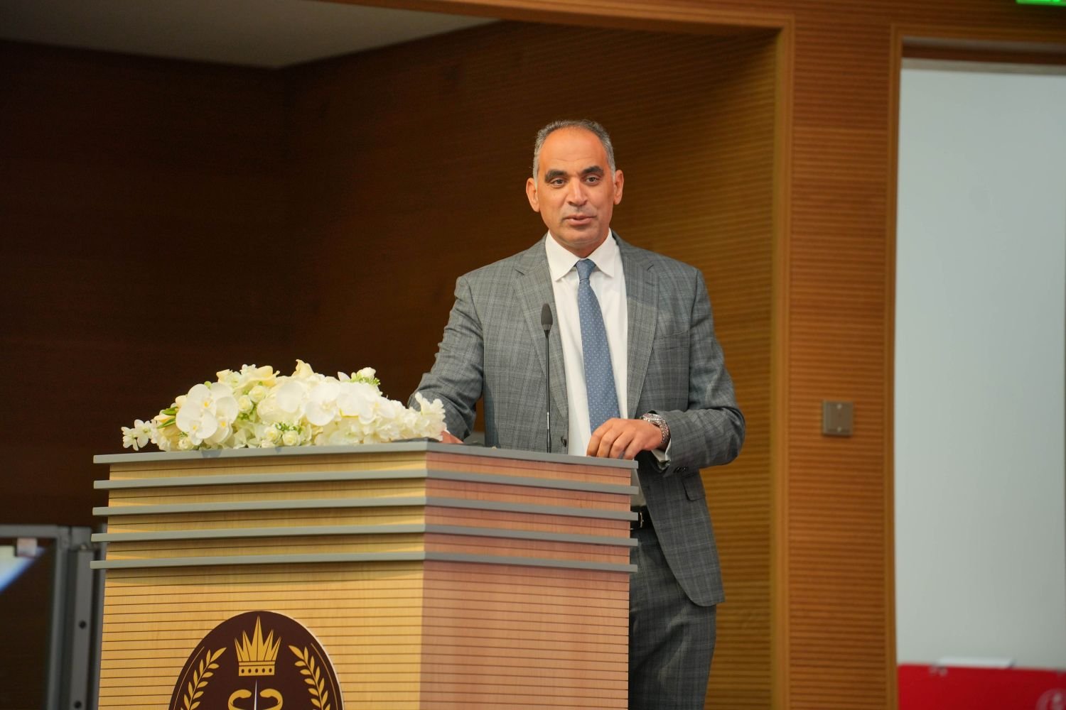
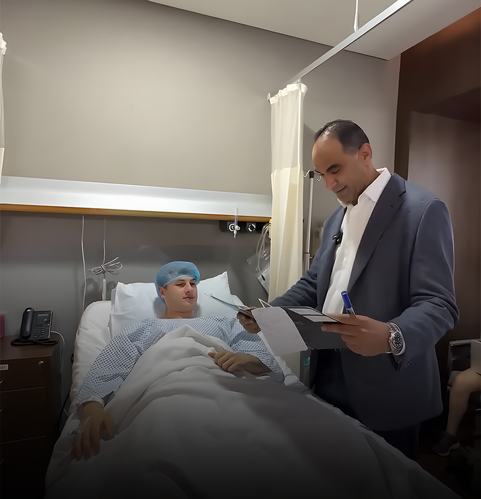
Dr. Usama Hassan Saleh

MD, PhD, MRCS (UK) | Egyptian & Arab Board Certified

Fellowship: Orthopedic Surgery, University of Toronto, Canada

With 23 years of orthopedic surgical practice in Dubai and the UAE, Dr. Usama is a specialist in knee ligament injuries, multi-ligament reconstruction, and complex knee instability management at Medcare MOSH.

23 Yrs
Expert Experience
Toronto
Fellowship Trained
Injury Overview

What Are Knee Ligament Injuries?

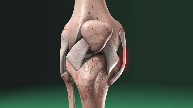
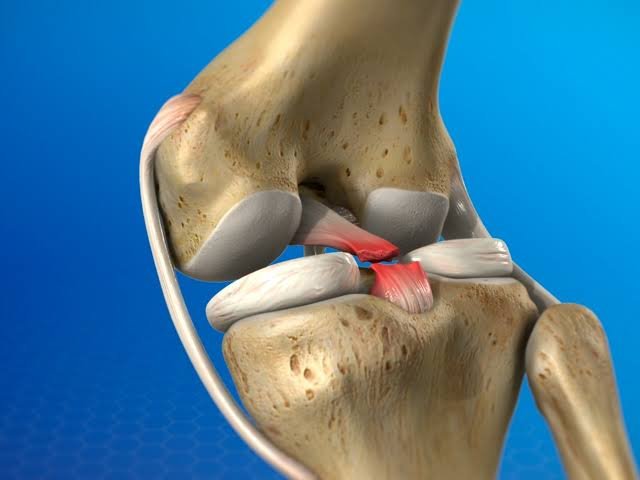
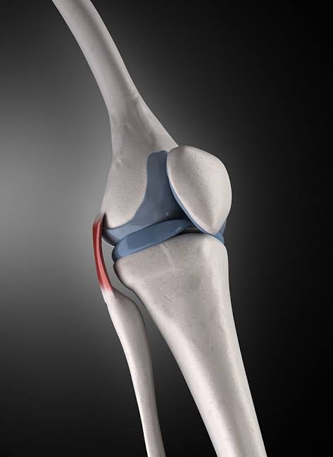
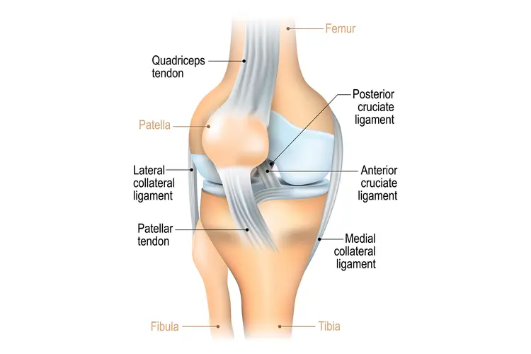
The knee is stabilized by four primary ligaments. The anterior cruciate ligament (ACL) and posterior cruciate ligament (PCL) cross inside the joint, controlling forward and backward tibial translation.

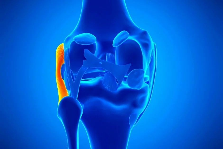
The medial collateral ligament (MCL) runs along the inner side of the knee, resisting valgus (inward) stress. The lateral collateral ligament (LCL) runs along the outer side, resisting varus (outward) stress. Together, they provide the stability required for normal knee function across all movement planes.
01

MCL Injuries

The most commonly injured ligament (~40%). Extra-articular location gives it excellent inherent healing capacity, allowing the vast majority of isolated tears to heal successfully without surgery.

02

Non-ACL Spectrum

This section covers the full clinical spectrum of non-ACL injuries: targeted pathways for complex PCL tears, isolated or combined MCL tears, LCL damage, and posterolateral corner injuries.

03

Multi-Ligament Cases

Complex high-impact trauma cases where two or more ligaments are simultaneously damaged. Each requires a distinct injury mechanism analysis, diagnostic examination, and surgical philosophy.

Treatment Philosophy

Understanding which ligament is injured and to what degree determines whether a patient needs structured home rehabilitation, physiotherapy, a hinged brace, or complex surgical reconstruction.

PCL Tears MCL Tears LCL Injuries Posterolateral Corner Multi-Ligament Damage Complex Knee Instability
Clinical Classifications

Types of Knee Ligament Injuries

Each knee ligament has a distinct injury mechanism, clinical presentation, and targeted treatment pathway.
MCL Tear (Medial Collateral Ligament) medical illustration
Most Common

MCL Tear (Medial Collateral Ligament)

Mechanism: Valgus force to the outer knee, forcing the inner knee to gap open (football/rugby tackle, late ski binding release, padel valgus collapse).

Graded 1-3. Produces medial (inner) knee pain, tenderness, and pain with valgus stress testing. Grade 1-2 and most grade 3 isolated MCL tears heal without surgery.
Recovery Time: 2 weeks (Grade 1) to 12 weeks (Grade 3)
PCL Tear (Posterior Cruciate Ligament) diagnostic visual
Strongest Ligament

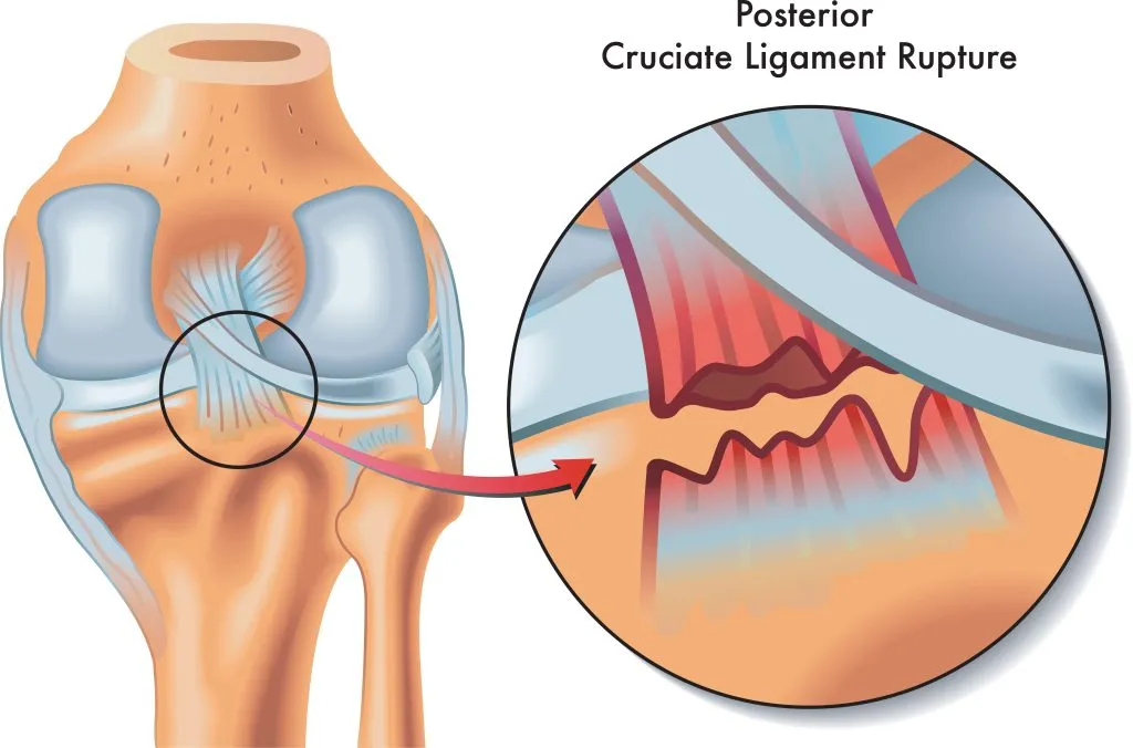
PCL Tear (Posterior Cruciate Ligament)

Mechanism: Direct posterior force to the tibia with the knee flexed (dashboard mechanism in collisions, blow from behind, or falling onto a bent knee).

Less common than ACL tears. Produces posterior knee pain, a positive posterior sag sign, and instability on stairs. Grade 1-2 are typically managed non-surgically.
Severe Cases: Grade 3 with multi-ligament requires surgery
LCL Tear (Lateral Collateral Ligament) trauma scan
Complex Matrix

LCL Tear (Lateral Collateral Ligament)

Mechanism: Varus force to the inner knee, forcing the outer knee to gap. Less common in isolation than MCL tears.

Often injured with posterolateral corner (PLC) structures. Produces lateral knee instability and may involve the peroneal nerve (foot dorsiflexion weakness).
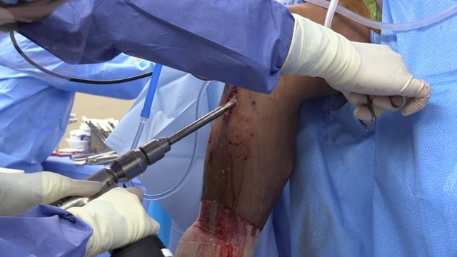
Surgical Indicator: Grade 3 due to poor lateral structure healing
Multi-Ligament Knee Injury high-energy trauma presentation
Urgent Case

Multi-Ligament Knee Injury

Mechanism: Simultaneous injury to two or more ligaments from high-energy trauma (motor vehicle collisions, heavy contact impacts, falls from height).

Knee dislocations are severe, with popliteal artery injury potential in 15-40%. Requires urgent neurovascular assessment and systematic MRI staging before surgery.

Pathway: Staged acute care + planned reconstructive surgery
Symptom Diagnostic Matrix

Clinical Signs & Symptoms

An advanced guide to evaluating knee ligament trauma by anatomical structure and injury grade severity.

Symptoms by Injury Grade

Clinical progression from micro-stretching to complete rupture.

01

Grade 1 Mild Sprain

Mild pain and tenderness at the injured ligament attachment points. Minimal swelling. The knee feels stable. May continue sports with discomfort. Tender to palpation directly over the ligament.

02

Grade 2 Partial Tear

Moderate pain, significant tenderness, and localized swelling. The knee may feel somewhat unstable during sport. Stress testing produces pain, but some restraint remains. 3-6 weeks of recovery.

03

Grade 3 Complete Tear

Complete disruption of the ligament. The knee feels clearly unstable; the injured side opens significantly on stress testing. Gross instability during weight-bearing, stair descent, and lateral movements.

Symptoms by Specific Ligament

Targeted indicators based on the exact damaged structure.

MCL
Medial Collateral Ligament

Medial (inner) knee pain and tenderness. Pain on valgus stress. Medial joint line bruising in grades 2-3. Point tenderness over the medial femoral epicondyle or medial tibial plateau attachment.

PCL
Posterior Cruciate Ligament

Posterior knee pain and pressure sensation. Difficulty and pain descending stairs and slopes. Positive posterior sag sign (tibia sags backward at 90° knee flexion). Significant functional limitation.

LCL
Lateral Collateral Ligament

Lateral (outer) knee pain and tenderness. Pain on varus stress. Lateral joint line bruising. Possible numbness or weakness of foot dorsiflexion (peroneal nerve involvement). Instability during varus loading.

CRITICAL
Multi-Ligament Injury

Gross instability in multiple planes. Major effusion developing rapidly. Inability to bear weight. Neurovascular warnings: absent pedal pulse (popliteal artery), foot drop (peroneal nerve). Requires emergency assessment.

Etiology & Risk Analysis

What Causes Knee Ligament Injuries?

Each knee ligament has a specific injury mechanism reflecting its anatomic function as a directional restraint.

MCL

MCL Mechanism Valgus Force

Valgus force to the outer knee when the foot is planted—a tackle from the side in football or rugby, a ski binding releasing late with the knee in valgus, or a lateral fall in padel or basketball. In Dubai's sporting population, football and rugby account for the majority of MCL injuries.

PCL

PCL Mechanism Direct Posterior Blow

Direct posterior tibial blow with the knee in flexion. The dashboard mechanism (knee striking the dashboard in a motor vehicle collision), a direct blow from behind in football, falling onto a flexed knee in rugby or martial arts, or hyperflexion in extreme sports.

LCL

LCL Mechanism Varus Force

Varus force to the inner knee, forcing the outer knee to gap. Less common than valgus injuries. LCL tears are more often associated with hyperextension injuries. Martial arts practitioners, cyclists, and high-speed impact sport athletes are at higher risk.

MULTI

Multi-Ligament Mechanism High-Energy Trauma

High-energy events: motor vehicle collisions, heavy contact sport impacts, and falls from height. Knee dislocation carries popliteal artery injury risk in up to 15-40% of complete dislocations. This is a vascular emergency.

Knee ligament stress diagnostics and mechanisms

Key Risk Factors

  • Contact and collision sports: rugby, football, basketball, martial arts
  • High-valgus loading activities: paddle, skiing, snowboarding
  • High-speed sport: road cycling, motorsport
  • Female athletes: higher rates of MCL injury in football and netball
  • Previous ipsilateral or contralateral knee ligament injury
  • Inadequate strength training: weak hip abductors & hamstrings increase valgus collapse risk
Clinical Diagnostics

Diagnosing PCL, MCL, & LCL Injuries

Accurate diagnosis requires targeted clinical examination using ligament-specific stress tests, confirmed with high-resolution imaging.

Targeted Clinical Examination

MCL

MCL Stress Testing

Valgus stress test at 0° (full extension, testing capsular integrity) and 30° (isolated MCL). Graded by millimeters of side-to-side opening: Grade 1 (<5 mm), Grade 2 (5-10 mm), Grade 3 (>10 mm).

PCL

PCL Clinical Assessment

Posterior drawer test (posterior force applied at 90°). Posterior sag sign where the tibia sags posteriorly under gravity at 90° flexion. Quadriceps active test to reduce posterior tibial sag.

LCL

LCL & PLC Testing Matrix

Varus stress test at 0° and 30°. External rotation test at 30° and 90° to implicate the posterolateral corner (PLC). Dial test utilized for combined LCL and PLC injury confirmation.

MULTI

Multi-Ligament Assessment

Full ligament examination plus urgent neurovascular assessment. Palpation of pedal pulses (dorsalis pedis and posterior tibial). Immediate assessment of foot dorsiflexion and sensation (peroneal nerve).
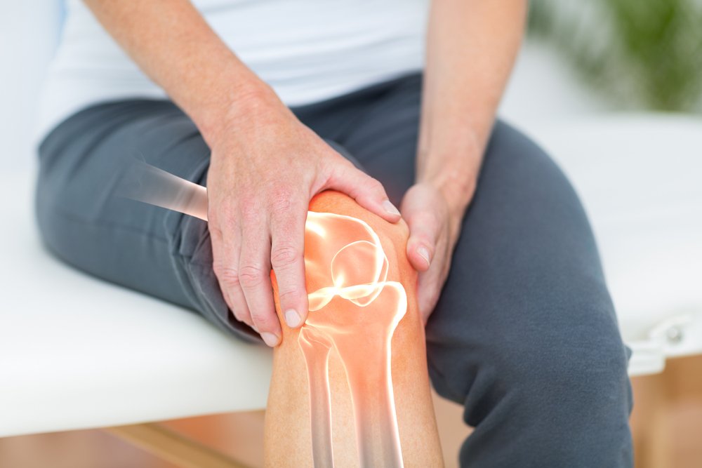
Dr. Usama Saleh performing advanced clinical diagnostic assessment

Advanced MRI Diagnostics Gold Standard

Confirms ligament grade, identifies bony avulsion, assesses coexisting meniscal and chondral damage (present in up to 50% of multi-ligament injuries), and guides precise surgical planning.

CT Angiogram Vascular Emergency

Urgently required for any suspected multi-ligament knee injury or knee dislocation to exclude popliteal artery injury before other investigations proceed. Popliteal artery injury is a surgical emergency.

Symptom Radar

Knee Ligament Symptoms

Recognising symptoms early by injury grade and specific structural location helps determine whether a patient needs structured rehab, bracing, or surgical reconstruction.

01

Grade 1 (Sprain)

Mild pain and tenderness at the injured ligament attachment points. Minimal swelling. The knee feels stable. May continue sports with discomfort. Tender to palpation directly over the ligament.

02

Grade 2 (Partial Tear)

Moderate pain, significant tenderness, and localized swelling. The knee may feel somewhat unstable during sport. Stress testing produces pain, but some restraint remains. 3-6 weeks of recovery.

03

Grade 3 (Complete Tear)

Complete disruption of the ligament. The knee feels clearly unstable, and the injured side opens significantly on stress testing. Gross instability during weight-bearing, stair descent, and lateral movements.

04

MCL Indicators

Medial (inner) knee pain and tenderness. Pain on valgus stress. Medial joint line bruising in grades 2-3. Point tenderness over the medial femoral epicondyle or medial tibial plateau attachment.

05

PCL Indicators

Posterior knee pain and pressure sensation. Difficulty and pain descending stairs and slopes. Positive posterior sag sign (tibia sags backward at 90 degrees knee flexion). Significant functional limitation.

06

LCL & Multi-Ligament

Lateral (outer) knee pain and tenderness. Pain on varus stress. Multi-ligament injuries produce gross instability in multiple planes, major effusion developing rapidly, and inability to bear weight.

Red Flag Indicators

When Should You See a Doctor in Dubai?

Accurate diagnosis of which ligament is injured and to what grade is the single most important step. Self-diagnosis is not a substitute for expert clinical examination and high-resolution imaging.

Immediate Emergency Assessment Required

Critical Alert

Gross Instability & Vascular Risk

CAUTION: Your knee is grossly unstable in multiple directions after a high-energy injury—this may indicate a knee dislocation with potential popliteal artery involvement. Popliteal artery injury requires surgical restoration of blood flow within 6-8 hours to prevent limb loss. Attend the nearest emergency department immediately. Do not wait for a scheduled appointment.

Nerve Involvement

Neurological Warning Signs

You have numbness, tingling, or weakness of the foot after a knee injury. Peroneal nerve injury from LCL and lateral knee dislocation requires prompt assessment to determine if nerve decompression is needed.

Seek Urgent Assessment (Within 24-48 Hours)

01

Rapid Hemarthrosis / Swelling

Your knee swelled significantly within 1-2 hours of a contact injury. Rapid hemarthrosis after contact injury may indicate cruciate or collateral ligament disruption beyond a simple sprain.

02

Functional Giving Way

Your knee feels clearly unstable—giving way during weight-bearing, stair descent, or lateral movements. Significant instability indicates Grade 3 ligament damage requiring imaging and specialist assessment.

Specialist Consultation Pathways

Unsettled Sports Injury

Knee pain and swelling following a sports injury have not settled after 1-2 weeks of RICE management. Persistent symptoms suggest a ligament injury rather than a simple contusion.

Misdiagnosed Knee Sprain

You have been told you have a knee sprain, but the knee does not feel right and is limiting your sport. Collateral ligament injuries are frequently dismissed as sprains without adequate examination or imaging.

Safe Return to Contact Sport

You want to return to contact sport after a knee injury and need confirmation that the ligament has healed adequately. Premature return to contact sport after an incompletely healed MCL is the most common cause of re-injury.

Clinical Rehabilitation

Exercises for Knee Ligament Injuries

A targeted, phase-specific guide on what protocol is safe to execute and when to safely initiate progressive mechanical loading.

Absolute Clinical Precautions

CAUTION: Do NOT perform exercises if the knee has gross instability in multiple planes (multi-ligament injury) without medical clearance. Do NOT apply valgus or varus stress to the knee in the acute phase (weeks 0-3) of MCL and LCL tears. Do NOT perform pivoting or rotation exercises before clinical and imaging confirmation that the ligament has healed adequately. For multi-ligament injuries: all exercise rehabilitation must be supervised by a physiotherapist coordinated with Dr. Usama.

MCL Protocol

MCL Tear Rehabilitation Exercises (Weeks 1-6)

Quad sets (Weeks 1-2)

Sit with the leg straight, tighten the quadriceps, hold for 5 seconds, and release. 3 sets of 15. Prevents quadriceps atrophy without knee joint loading. Safe from day 1 of injury.

Straight leg raises (Weeks 1-3)

Lie on your back, affected leg straight, tighten your quad, and raise to 45 degrees; hold for 3 seconds and lower slowly. 3 sets of 15. Strengthens quad without MCL stress.

Stationary cycling (Weeks 2-4)

Low resistance, comfortable range of motion. Begin as soon as the brace allows 90 degrees of knee flexion. Maintains cardiovascular fitness without valgus loading.

Single-leg balance (Weeks 3-6)

Stand on the affected leg with a slight knee bend for 30 seconds, progressing to 60 seconds. Eyes open first, then eyes closed. Restores proprioception critical for sport return.

PCL Focus

PCL Rehabilitation Focus

Quadriceps strengthening is the absolute priority for PCL rehabilitation to counteract posterior subluxation.

Terminal Knee Extension (TKE)

Stand with a resistance band around the back of the knee and straighten the knee fully against resistance. 3 sets of 15. The single most important PCL rehabilitation exercise.

Contraindication Warning

Avoid hamstring-dominant exercises (leg curls, hamstring-dominant deadlifts) for the first 6-8 weeks—hamstrings produce a posterior tibial force that stresses the healing PCL.

LCL Focus

LCL Rehabilitation Focus

Hip Abductor Strengthening

Weak hip abductors increase varus collapse during single-leg activities, directly stressing the LCL. Side-lying hip abduction: raise the top leg to 45 degrees, hold for 3 seconds, and lower slowly. 3 sets of 15. Begin strictly from week 2.
Exercises to Avoid (Acute Phase)

  • Pivoting and cutting movements before absolute clinical clearance.
  • Valgus or varus loading activities (deep squatting with knee-in collapse).
  • Contact sport or high-impact jumping before stability confirmation.
  • Any exercise producing localized pain at the injured ligament site during loading.
Knee ligament clinical assessment
Clinical Diagnostics
Dr. Usama Saleh consultation
Expert Consultation
Knee joint rehabilitation training
Rehabilitation Guide
Advanced physical therapy
Targeted Exercise Recovery
Risk & Prognosis Timeline

What Happens if Left Untreated?

The consequences of untreated or inadequately managed knee ligament injuries depend entirely on the ligament and grade. Progression leads to severe functional and surgical complications.

01

Chronic Knee Instability

The most consistent consequence of untreated Grade 3 collateral ligament injuries. A chronically unstable knee gives way repeatedly during sports and daily activities. Each giving-way episode risks secondary meniscal tears and severe cartilage damage.

02

Progressive Meniscal Damage

An MCL-deficient or PCL-deficient knee subjects the menisci to abnormal shear and compression forces. Secondary meniscal tears develop at rates of approximately 10-15% per year in chronically ligament-deficient knees, accelerating cartilage loss further.

03

Osteoarthritis Acceleration

Combined ligament deficiency and meniscal damage produce a joint environment driving early-onset knee osteoarthritis (OA). Long-term clinical studies demonstrate significantly higher OA rates in patients with untreated Grade 3 collateral ligament injuries.

Critical

Vascular & Neurological Compromise

Popliteal artery injury in an unrecognized knee dislocation can result in lower limb ischemia, requiring emergency vascular surgery or risking limb loss. Delayed recognition is the leading cause of amputation. Peroneal nerve injury can result in permanent foot drop.

05

Increased Surgical Complexity with Delay

Grade 3 MCL and PCL injuries that develop chronic scar tissue, deformity, or secondary meniscal damage are significantly more complex to reconstruct than acute presentations. Early definitive management produces better outcomes with lower surgical risk than delayed reconstruction.

Patient Readiness

Preparing for Your Injury Assessment

A structured step-by-step roadmap to prepare for your MCL, PCL, or LCL assessment and surgical or non-surgical treatment pathways in Dubai.

Preparation Stage
Clinical Guidelines & Actions

Immediate Care

Acute Phase (RICE)

Rest (non-weight-bearing if unstable), Ice (20 minutes every 2-3 hours for the first 48-72 hours), Compression (elastic bandage), and Elevation (above heart level).

Avoid NSAIDs for the first 48-72 hours if surgical intervention is possible. Please arrange crutches for suspected Grade 2-3 injuries.

Consultation

Clinical Evaluation

Please bring a comprehensive injury description (exact mechanism, what you heard or felt at the moment of impact, and how quickly the swelling developed).

  • Bring all previous knee imaging (X-Rays, MRI scans) and a list of current medications.
  • Share your specific sport and athletic training demands with the team.
  • Dr. Usama performs structural stress testing at consultation—wear shorts for easy knee access.

Pre-Surgical

Prehab & Readiness

Engage in prehab physiotherapy for 4-6 weeks where time allows to optimize post-operative recovery outcomes. Complete smoking cessation is required a minimum of 4 weeks before surgery.

Stop all blood-thinning medications per specific medical instructions. Arrange home preparation for a crutch-dependent recovery during the first 4-6 weeks post-op.

Surgical Orchestration

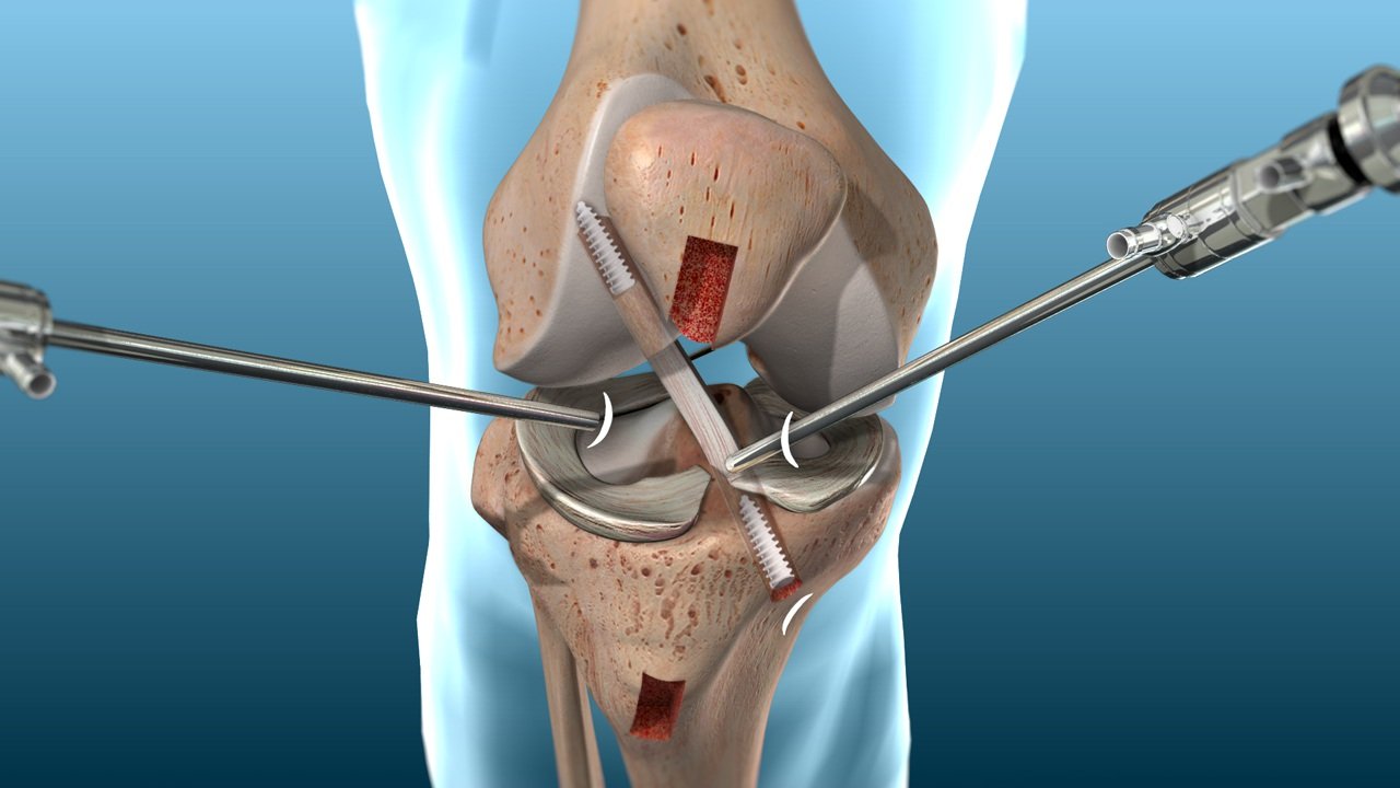
How MCL Reconstruction Is Performed

A comprehensive anatomical guide detailing the precise step-by-step surgical execution and postoperative recovery specifications in Dubai.

15-20 Mins

Anesthesia and Positioning

Administered via general anesthesia paired with a targeted femoral nerve block for advanced postoperative pain control. The patient is positioned supine with a tourniquet applied, followed by full stabilization, preparation, and sterilization of the surgical field.

10-15 Mins

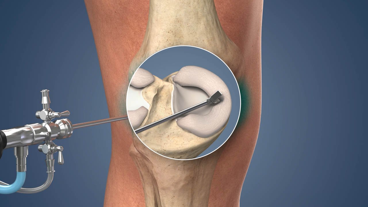
Arthroscopic Assessment

Comprehensive arthroscopic examination of the knee joint interior. This vital phase confirms any coexisting meniscal or cartilage pathology, thoroughly assesses ACL and PCL status, and completely verifies the intra-articular injury blueprint before open reconstruction begins.

10-15 Mins

Graft Preparation

The structural graft is typically harvested from the ipsilateral gracilis tendon (via a precise 2-3 cm incision at the pes anserinus) or sourced using an advanced allograft tendon. The tissue is meticulously prepared to match the perfect anatomical diameter and length requirements.

30-45 Mins
Core Procedure

MCL Anatomic Reconstruction

An incision is executed directly over the medial femoral condyle. The native MCL remnant is carefully identified and preserved wherever possible to protect intrinsic receptors. Anatomic attachment footprints are localized on both the femur and tibia. The prepared graft is then locked securely at these femoral and tibial attachment sites using high-grade interference screws or cortical buttons, successfully restoring the original MCL isometric line.

10-15 Mins

Closure and Brace Application

Layered closure of the medial capsule and supporting soft tissues. A functional hinged knee brace is applied directly in the operating theater, locked securely between 30-60 degrees of flexion for the first 2 weeks to safely insulate the healing graft matrix.

Procedure Specifications

Total Surgical Time
75-90 Minutes

For isolated MCL reconstructions.

Hospital Discharge
Same-Day or Next Morning

Dependent upon final case complexity.

Crutches Required
4-6 Weeks

Postoperatively to manage early loading.

Physiotherapy Protocol
Week 1-2 Post-Surgery

Early motion under controlled activation.

Return to Contact Sport
6-9 Months

Following full remodeling confirmation.

Prognosis & Return Pathways

Recovery After Ligament Treatment

A comprehensive clinical guide breaking down surgical and non-surgical recovery tracking parameters across all knee injury classifications in Dubai.

MCL Injury

Grade 1 (Mild Sprain)

Non-Surgical Pathway: 1-3 Weeks
Return to Sport: 1-3 Weeks
MCL Injury

Grade 2 (Partial Tear)

Non-Surgical Pathway: 3-6 Weeks
Return to Sport: 4-6 Weeks
MCL Injury

Grade 3 (Non-Surgical)

Non-Surgical Pathway: 6-12 Weeks
Return to Contact Sport: 6-12 Weeks
Surgical Pathway

Grade 3 (Reconstruction)

Surgical Recovery: 6-9 Months
Return to Contact Sport: 6-9 Months
PCL Injury

Grade 1-2 (Cruciate Sprain)

Non-Surgical Pathway: 4-8 Weeks
Return to Sport: 4-8 Weeks
Surgical Pathway

PCL Grade 3 (Severe Tear)

Surgical Recovery: 9-12 Months
Return to Sport: 9-12 Months
Surgical Pathway

LCL Grade 3 (Lateral Complex)

Surgical Recovery: 6-9 Months
Return to Sport: 6-9 Months
High-Energy Trauma

Multi-Ligament (Surgical)

Surgical Recovery: 12-18 Months
Return to Contact Sport: 12-18 Months

Mandatory Return-To-Sport Safety Criteria

No patient should return to contact sport based on pain resolution alone without proper structural stability confirmation from a specialist.

Ligament Stability: Absolute restriction check passed and confirmed via clinical objective stress testing.

Muscular Strength Symmetry: Quadriceps and hamstring strength testing scores strictly above 90% of the contralateral healthy limb.

Functional Stability: Single-leg hop test mechanical symmetry tracking scores validated above 90% accuracy.

Agility Readiness: Full clearance achieved and validated across sport-specific functional agility testing matrices.

Clinical Prognosis

Risks & Expected Outcomes by Ligament Type

Statistical success indicators, complication risk parameters, and functional athletic return rates validated across worldwide orthopedic registries.

95-100%
MCL Grade 1-2

Non-Surgical Pathway Return

Achieves excellent functional restoration. Clinical data proves that the structural re-injury rate is significantly reduced with progressive, physiotherapy-confirmed healing protocols before sport return.

80-90%
MCL Grade 3

Non-Surgical Contact Sport Return

Validated at the 3-month mark post-injury, heavily contingent upon strict hinged brace compliance and intensive, progressive resistance physical therapy orchestration.

85-90%
MCL Surgery

Anatomic Reconstruction Success

Expected return to previous high-demand sport within 6-9 months. Risk matrices indicate an exceptionally low infection rate of less than 1%, and a graft failure safety index below 5% globally.

75-85%
PCL Surgery

Cruciate Reconstruction Profile

Successful return to pre-injury sports levels. Orthopedic metrics confirm the best clinical outcomes are achieved only with a strict, intensive quadriceps strengthening isolation protocol.

80-85%
LCL & PLC Surgery

Lateral Restraint Remodeling

Return to sports proficiency. Peroneal nerve functional numbness or weakness typically improves with prompt structural decompression. Outcomes are significantly worse when reconstruction is delayed beyond 3 months.

60-75%
Complex Trauma

Multi-Ligament Global Return

Return to previous competitive sport. Outcomes remain highly dependent upon the initial injury velocity severity, acute vascular status, nerve footprint baseline, and absolute rehabilitation completeness.

Why Choose Us

Why Choose Dr. Usama for Knee Ligament Injuries?

Two decades treating Dubai's competitive sporting community. Dr. Usama provides subspecialty, fellowship-level expertise from diagnosis to complex multi-ligament surgical reconstruction under one roof.

Global Expertise. Local Heart.
MD PhD MRCS (UK) 23 Yrs Experience Fellowship-Trained (Canada)

Fellowship-Trained Knee Specialist at Medcare MOSH

01

Fellowship-Level Knee Expertise

Advanced fellowship training at the University of Toronto provided subspecialty expertise in multi-ligament knee reconstruction, PCL surgery, and posterolateral corner reconstruction—procedures requiring specific fellowship-level training well beyond standard orthopedic qualifications.

02

Accurate Structural Diagnosis

MCL, PCL, LCL, and PLC injuries produce completely different clinical signs and require different treatment pathways. Dr. Usama uses validated clinical tests for each individual ligament, confirmed via high-resolution MRI, preventing generic or inaccurate knee diagnoses.

03

Conservative-First Philosophy

Surgery is never the default option. The vast majority of MCL tears and many PCL tears heal successfully without surgery. Dr. Usama avoids surgical risk by preserving biological tissue, ensuring equivalent outcomes for Grade 1-2 collateral injuries through structured rehab.

04

Full-Spectrum Care at Medcare MOSH

From same-consultation dynamic stress testing and MRI review to hinged brace fitting, advanced physiotherapy coordination, and complex surgery—all executed seamlessly at Medcare Orthopaedics and Spine Hospital on Sheikh Zayed Road, Dubai.

05

Complex Multi-Ligament Capability

High-stakes multi-ligament injuries require advanced surgical planning. Dr. Usama's subspecialty background covers the staged architectural approach to multi-ligament reconstruction, making Medcare MOSH one of the few centers with full complex knee capability.

06

23 Years of Dubai Sporting Experience

Two decades treating Dubai's football, rugby, padel, martial arts, and cycling athletes. Deep familiarity with the specific knee ligament injury patterns of Dubai's sporting community means faster recovery protocols calibrated directly to your activity goals.
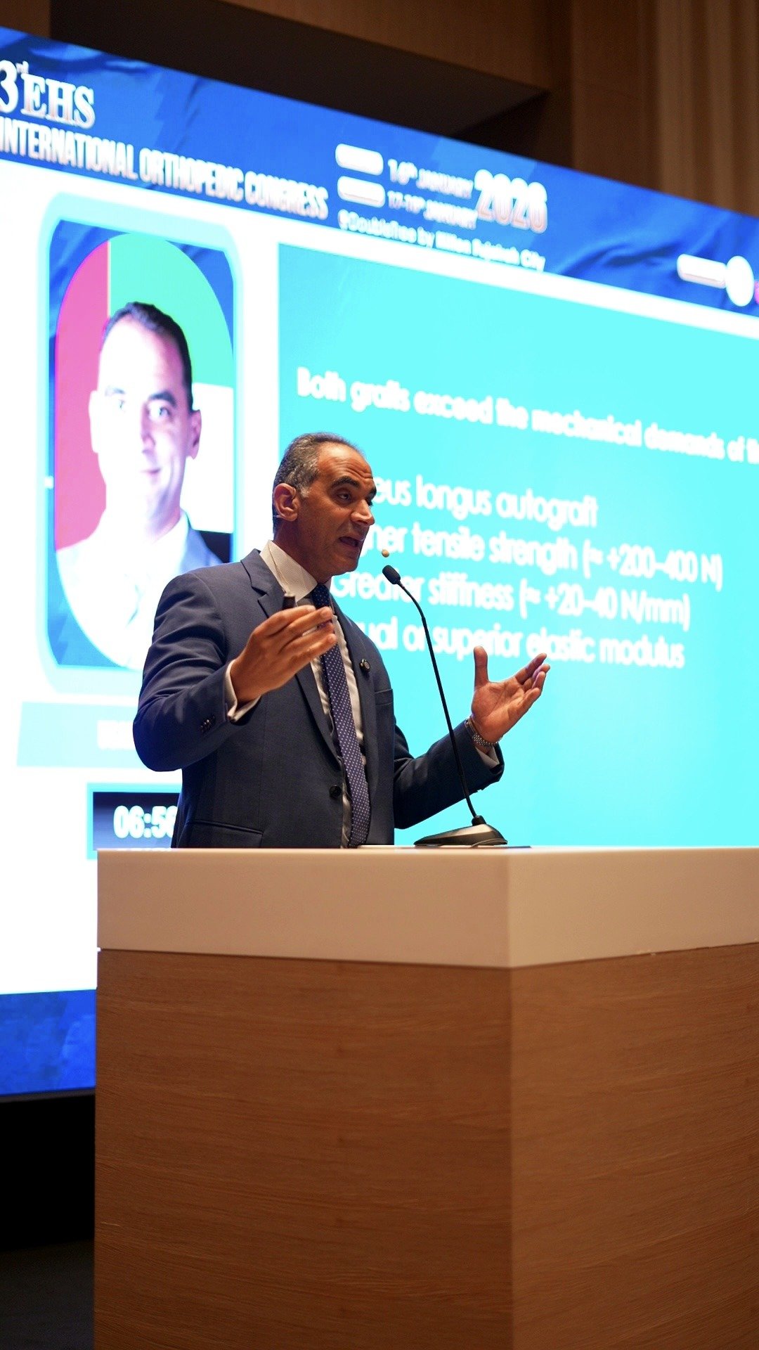
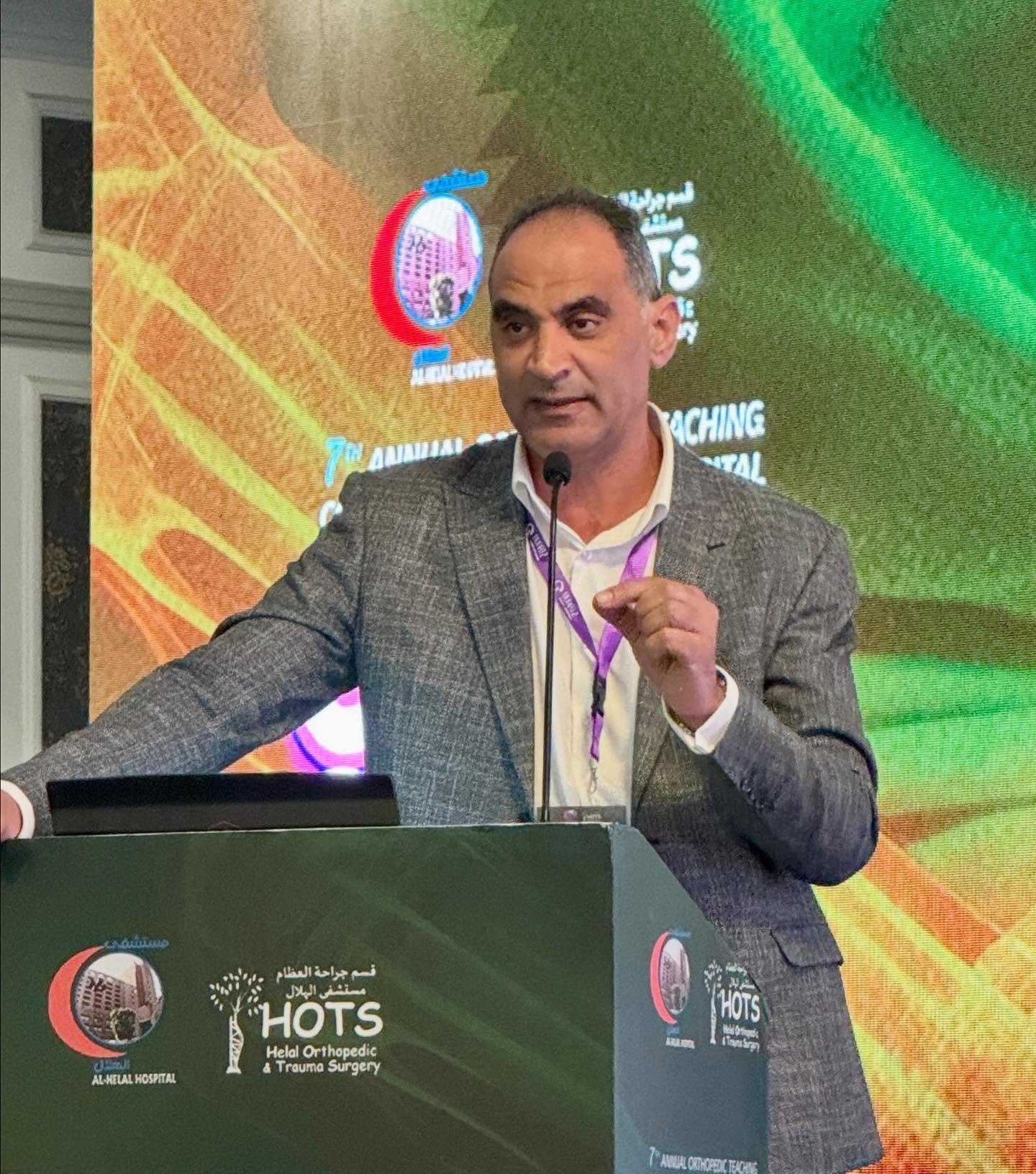
Diagnostics 1
Diagnostics 2
Diagnostics 3
Diagnostics 4
Diagnostics 1
Diagnostics 2
Diagnostics 3
Diagnostics 4
Diagnostics 5
Diagnostics 6
Diagnostics 7
Diagnostics 8
Diagnostics 5
Diagnostics 6
Diagnostics 7
Diagnostics 8
UAE Sports Care Network

Knee Ligament Injury Treatment in Dubai

Serving competitive athletes across the UAE. From local football and rugby clubs to padel and high-speed sports cycling, Medcare MOSH delivers centralized, world-class orthopedic diagnostic and surgical orchestration.

Targeted Sports Profiles

Dubai's contact and collision sports culture produces a high-volume demand for knee ligament assessments. Dr. Usama regularly treats rugby players from Dubai Exiles and Hurricanes, football players from Al Quoz to Jumeirah leagues, padel athletes from JBR to Business Bay, and high-speed cycling injuries.

Geographic Patient Reach

Dr. Usama sees knee ligament injury patients from all major Dubai communities including Jumeirah, Dubai Marina, JBR, Downtown, Business Bay, and Al Barsha. Due to his subspecialty expertise, patients also regularly travel from Abu Dhabi, Ajman, and across the northern UAE.

Primary Clinical Facility

Medcare Orthopaedics and Spine Hospital (MOSH)
Located centrally on Sheikh Zayed Road, Dubai, UAE. This flagship facility provides seamless, single-station access to advanced dynamic stress testing, high-resolution MRI imaging, and state-of-the-art orthopedic operating theaters.

Why Choose Dr. Usama
For Shoulder Impingement Treatment In Dubai

Best Shoulder Doctor Dubai — Fellowship-Trained

23+ Years as Dubai's Shoulder Specialist Doctor

Expert in How to Treat Rotator Cuff Tear — All Options

Honest — Surgery Only When Truly Needed

Patient Knowledge Base

Frequently Asked Questions

Expert clinical answers regarding PCL, MCL, and LCL injuries, recovery timelines, and treatment pathways in Dubai.

MCL tear recovery time depends on the grade of injury. Grade 1 MCL sprain: 1-3 weeks to return to normal activity and sport with appropriate management. Grade 2 MCL partial tear: 3-6 weeks to full sport, often with a hinged brace for the first 4 weeks. Grade 3 MCL complete tear (non-surgical): 6-12 weeks to return to contact sport with intensive physiotherapy and brace compliance. Grade 3 MCL requiring surgical reconstruction (combined with ACL or refractory instability): 6-9 months to return to sport. The key variable in MCL tear recovery time is confirming adequate ligament healing on clinical examination before return to contact sport, not simply waiting for pain to settle.

Yes—the vast majority of MCL tears heal successfully without surgery. The MCL has excellent biological healing capacity because of its extra-articular location and blood supply. Grade 1 and Grade 2 MCL tears: non-surgical management (rest, brace, and physiotherapy) produces a 95-100% return to pre-injury activity. Grade 3 isolated MCL tears: 80-90% heal adequately with a hinged brace and intensive physiotherapy over 6-12 weeks. Surgery for isolated MCL tears is reserved for refractory instability at 3 months of conservative management or when the MCL is torn simultaneously with the ACL or PCL.

Symptoms depend on which ligament is injured. Common features: pain at the time of injury (a pop or crack most common with Grade 3 tears), swelling (haemarthrosis developing within 1-2 hours for major tears), and instability (the knee feeling loose or giving way). Ligament-specific: MCL -- inner knee pain and tenderness, pain on valgus stress. PCL—posterior knee pain, instability on stairs and slopes. LCL—outer knee pain, possible foot numbness or weakness (peroneal nerve). Multi-ligament—gross instability, major effusion, absent pedal pulse (emergency). Any knee injury producing immediate swelling and instability requires specialist assessment and MRI within 48-72 hours.

The MCL (medial collateral ligament) runs along the inner side of the knee and is injured by a valgus (inward) force—typically a contact injury to the outer knee. MCL tears are extra-articular, have a good blood supply, and the majority heal without surgery. Recovery: 2-12 weeks depending on grade. The ACL (anterior cruciate ligament) runs through the center of the knee and is typically injured by a non-contact pivoting or hyperextension mechanism. ACL tears are intra-articular, have no blood supply for self-healing, and active patients requiring return to pivoting sports need surgical reconstruction. Recovery after ACL reconstruction: 9-12 months. The two ligaments can be torn simultaneously in the unhappy triad injury (ACL, MCL, and medial meniscus), requiring comprehensive surgical planning.

Treatment depends entirely on which ligament is injured and the grade. Grade 1-2 collateral ligament tears: The RICE protocol for 48-72 hours, a hinged brace for Grade 2, and physiotherapy-led rehabilitation over 3-6 weeks. Most do not require surgery. Grade 3 MCL: usually non-surgical—MCL tear recovery time is 6-12 weeks with bracing and intensive physiotherapy. Grade 3 LCL usually requires surgical reconstruction. PCL Grade 1-2: conservative management with quadriceps strengthening. PCL Grade 3 in active athletes: assessment dependent. Multi-ligament injuries: emergency neurovascular assessment and staged surgical reconstruction. The most important first step is accurate diagnosis: the correct ligament identified, graded accurately, and coexisting injuries assessed correctly.

A posterior cruciate ligament (PCL) tear produces posterior knee pain, a sense of the tibia sagging backward when the knee is bent, difficulty descending stairs and slopes, and a positive posterior sag sign and posterior drawer test on examination. Treatment: Grade 1-2 PCL tears are managed with quadriceps strengthening (the quads resist the posterior tibial sag that PCL deficiency produces), activity modification, and physiotherapy over 4-8 weeks with excellent outcomes. Grade 3 PCL tears in active athletes with functional instability, or combined with other ligament injuries, typically require PCL reconstruction followed by 9-12 months of rehabilitation.

Knee ligament surgery costs in Dubai: MCL reconstruction AED 30,000-50,000; PCL reconstruction AED 40,000-60,000; LCL and ACL reconstruction AED 40,000-65,000; and multi-ligament reconstruction AED 60,000-100,000 depending on complexity. Consultation and MRI: AED 400-800 (consultation) plus AED 1,200-2,500 (MRI). Most major UAE insurance plans—Daman, DHA, Cigna, AXA, Bupa, MetLife, GlobeMed, and NAS—cover medically necessary knee ligament reconstruction for confirmed Grade 3 injuries and surgical candidates. Dr. Usama's team at Medcare MOSH verifies coverage and provides transparent cost estimates before any procedure.

Dr. Usama Saleh at Medcare Orthopaedics and Spine Hospital (MOSH), Sheikh Zayed Road, Dubai, is a fellowship-trained orthopedic surgeon with 23 years of knee ligament injury experience covering isolated Grade 1 MCL sprains to complex multi-ligament knee reconstructions. His fellowship training at the University of Toronto provided subspecialty expertise in ligament surgery beyond standard orthopedic qualifications. If you are searching for a knee instability specialist near me in Dubai or an MCL specialist near you, Dr. Usama's team at Medcare MOSH can confirm same-day or urgent appointment availability.

Take control of your health, mobility, and quality of life.

Your First Step Toward a Stronger Tomorrow Starts Now:

Book your personal consultation with Dr. Usama Saleh today and begin your journey to a better you.

Contact Form dr

Powered by ICONCLY Medical.

Copyright © 2026. All rights reserved.

Patient Connection Hub Assess your pain