Plantar Fasciitis Treatment: Complete Relief & Recovery Guide
Plantar Fasciitis Treatment: Complete Clinical Guide to Heel Pain Relief Clinically reviewed by the orthopedic and sports injury team to provide evidence-based...
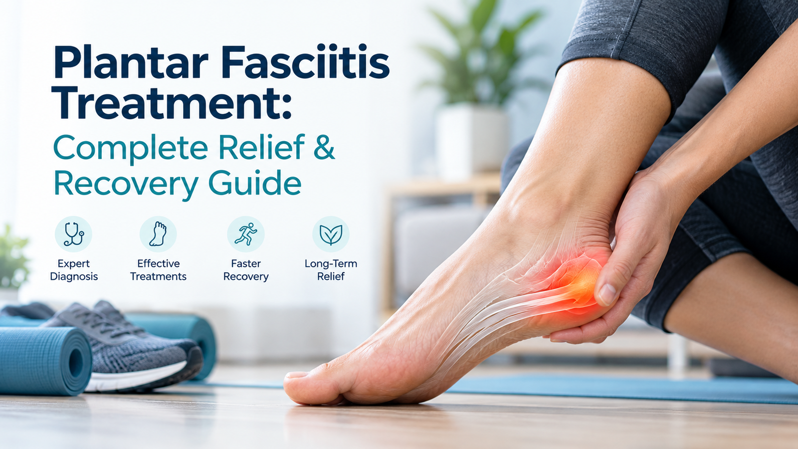
Plantar Fasciitis Treatment: Complete Clinical Guide to Heel Pain Relief
Clinically reviewed by the orthopedic and sports injury team to provide evidence-based recovery protocols for acute and chronic heel pain.
Sharp morning heel pain makes finding an effective plantar fasciitis treatment essential for restoring daily mobility. This condition occurs when the thick band of tissue supporting your foot arch becomes irritated from repetitive strain. Understanding proven non-surgical therapies, targeted strengthening exercises, and advanced clinical options helps you resolve discomfort and protect long-term foot biomechanics without unnecessary delays.
Understanding Plantar Fasciitis: Anatomy, Biomechanics, and First-Step Pain
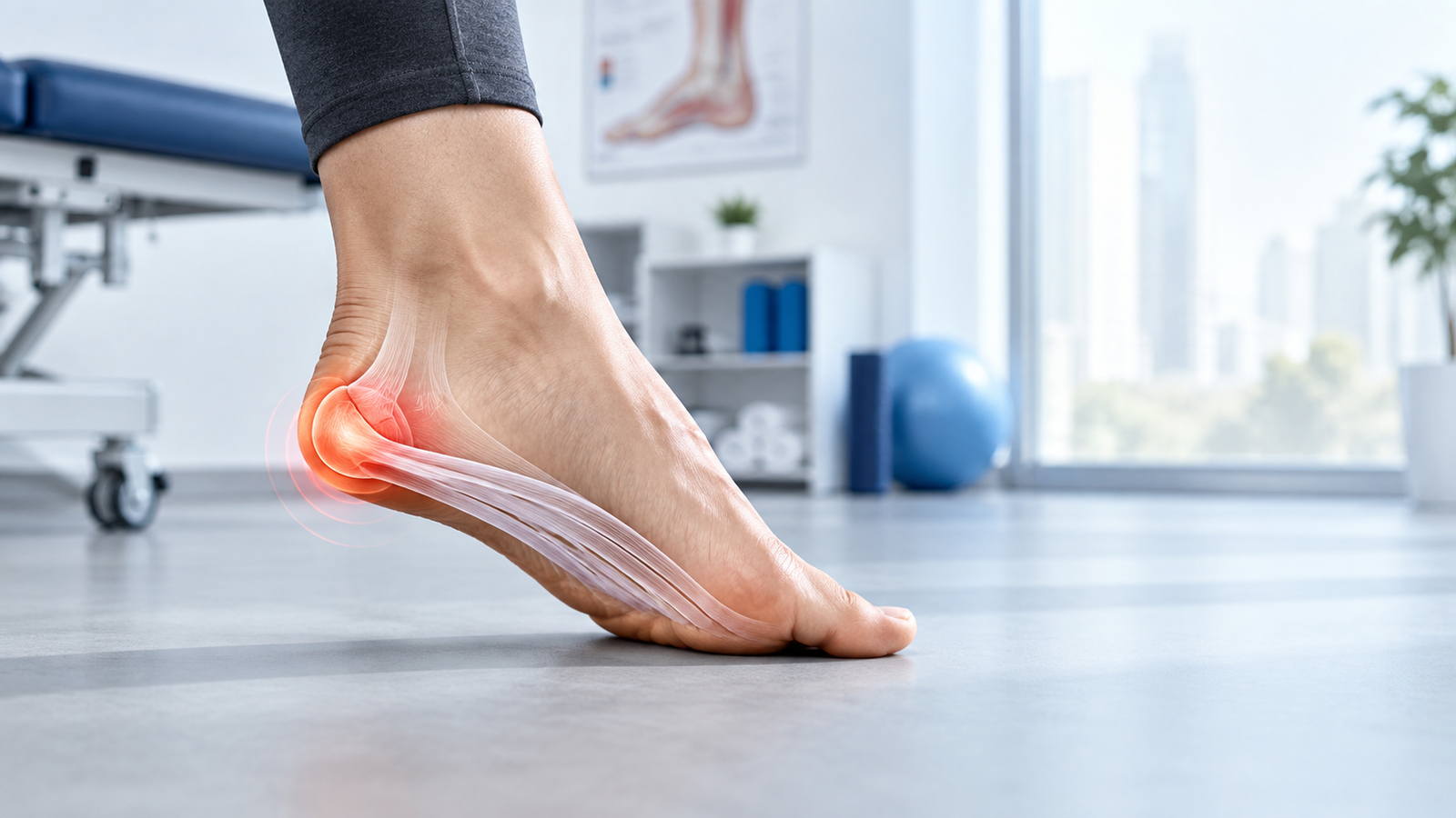
The plantar fascia is a dense band of fibrous connective tissue running along the bottom of your foot. It connects your heel bone (calcaneus) to the base of your toes.
This structure functions like a shock-absorbing bowstring, supporting your foot arch and distributing mechanical tension during movement. When excessive load creates micro-tears faster than the body can repair them, localized degeneration develops.
Clinical practice highlights that chronic cases involve collagen disorganization (fasciosis) rather than simple inflammatory swelling.
Why Is Heel Pain Worst During the First Steps in the Morning?
During overnight sleep, your foot rests in a relaxed, downward position known as plantarflexion. This posture allows the healing plantar fascia fibers to shorten and tighten.
Stepping out of bed forces the foot into sudden dorsiflexion, placing immediate full body weight onto the contracted tissue. This sudden mechanical stretch pulls sharply on the calcaneal attachment site.
As you walk, the tissue warms up and elongates, temporarily reducing pain until prolonged weight-bearing triggers strain again.
Phase 1: Immediate Pain Reduction and First-Line Conservative Care
Initial management focuses on offloading strained tissue, reducing acute sensitivity, and preventing continuous micro-trauma. Starting early prevents chronic structural changes.
Most patients achieve meaningful relief by combining targeted daily home habits:
- Activity Modification: Replace high-impact running or jumping with low-impact swimming or cycling to decrease mechanical load.
- Ice Bottle Massage: Roll the arch of your foot over a frozen water bottle for 10 to 15 minutes twice daily to calm irritated nerve endings.
- Night Splints: Wear an adjustable tension splint to hold the foot at a neutral 90-degree angle overnight, preventing morning contraction.
- Supportive Indoor Footwear: Avoid walking barefoot on hard tile or hardwood floors, especially upon waking.
How Long Does It Take for Plantar Fasciitis to Go Away with Rest and Ice?
Mild acute cases often demonstrate measurable symptom reduction within four to six weeks of diligent conservative care. Complete recovery, however, requires structured tissue adaptation.
Relying solely on rest without addressing tendon load capacity frequently leads to symptom recurrence once normal activity resumes. You can evaluate your symptoms early using our interactive online orthopedic pain assessment quiz tool to gauge your current recovery stage.
Phase 2: Evidence-Based Physical Therapy and High-Load Strength Training
Modern sports medicine prioritizes active tissue remodeling over prolonged rest. Tendons and fascial bands adapt positively to progressive, controlled mechanical tension.
Physical rehabilitation should combine specific calf lengthening with progressive tensile loading protocols.
High-Load Strength Training vs. Passive Stretching: The Rathleff Protocol
While passive gastrocnemius stretching offers short-term comfort, high-load resistance training promotes collagen reorganization and increases fascial load tolerance.
The Rathleff protocol utilizes controlled heel raises performed on an elevated step with a rolled towel under the toes:
- Execution: Stand on the edge of a step with a rolled towel placed under your toes to maximize the windlass mechanism.
- Tempo: Rise upward over 3 seconds, hold at the top for 2 seconds, and lower smoothly over 3 seconds.
- Dosage: Perform 3 sets of 12 repetitions every other day, gradually adding backpack weight as strength improves over 12 weeks.
Phase 3: Orthotics, Footwear Mechanics, and Biomechanical Support
Foot structure strongly influences fascial strain. Both flat feet (overpronation) and rigid high arches (pes cavus) alter force distribution through the kinetic chain.
Selecting appropriate mechanical supports helps offload the medial calcaneal tubercle during daily standing and walking.
Custom Orthotics vs. Over-the-Counter Inserts: What Works Best?
Clinical trials show that both prefabricated and custom orthotics provide substantial pain relief by supporting the medial longitudinal arch.
The table below summarizes key differences between common supportive modalities:
| Support Modality | Primary Mechanism | Clinical Indication | Expected Durability |
| Prefabricated Firm Arch Supports | Reduces arch collapse and peak plantar pressure | Mild to moderate acute heel pain; neutral foot types | 6 to 12 months |
| Custom Functional Orthotics | Corrects complex rearfoot eversion and forefoot varus | Severe biomechanical abnormalities or recurrent symptoms | 2 to 5 years |
| Silicone Heel Cups | Cushions vertical heel strike impacts | Isolated fat pad thinning or localized heel bruising | 3 to 6 months |
| Rocker-Bottom Footwear | Facilitates smooth sagittal rollover, reducing toe dorsiflexion | Persistent morning stiffness and forefoot-driven fascial pull | 300 to 500 miles |
Patients managing persistent discomfort alongside broader lower-limb conditions often explore effective non-surgical orthopedic treatments for pain relief to balance their overall joint alignment.
Phase 4: Advanced Clinical Interventions for Persistent Heel Pain
When structured conservative care for 8 to 12 weeks yields insufficient improvement, advanced orthopedic interventions should be considered.
Non-invasive and minimally invasive clinical therapies target chronic degenerated tissue to stimulate an accelerated healing response.
Extracorporeal Shockwave Therapy (ESWT) vs. Corticosteroid Injections
Extracorporeal Shockwave Therapy delivers acoustic energy pulses to the plantar fascia insertion site. This process promotes microvascular ingrowth and downregulates pain receptors without invasive tissue disruption.
Conversely, while corticosteroid injections deliver powerful short-term anti-inflammatory relief, repeated injections carry documented risks:
- Plantar Fascia Rupture: Steroids can weaken the structural collagen matrix, increasing spontaneous rupture risk.
- Fat Pad Atrophy: Injection into the subcalcaneal fat pad can cause permanent cushion loss, creating intractable heel impact pain.
- ESWT Advantage: Shockwave therapy carries no risk of tissue thinning and supports long-term structural integrity.
Biological and Platelet-Rich Plasma (PRP) Injections
Autologous platelet-rich plasma (PRP) uses concentrated growth factors from your own blood to stimulate cellular regeneration in chronic fasciosis.
Specialized centers frequently utilize advanced biological repair and regenerative platelet therapies to treat stubborn connective tissue degeneration safely.
Choosing the Right Clinical Plan for Plantar Fasciitis Treatment
Navigating modern medical interventions requires matching each therapy to symptom chronicity and tissue quality. When basic conservative methods plateau, combining progressive mechanical loading with regenerative therapies offers a targeted plantar fasciitis treatment plan that rebuilds cellular health without prolonged downtime.
2026 Clinical Updates: Modern Shifts in Plantar Fascia Tissue Preservation
Recent orthopedic consensus guidelines updated through September 2026 emphasize tissue preservation over aggressive steroid ablations or early surgical release.
Key clinical shifts in contemporary practice include:
- Ultrasound-Guided Micro-Debridement: Minimally invasive percutaneous interventions now use focused ultrasonic energy to clear scarred tissue while sparing healthy fibers.
- Load-Management Over Complete Immobilization: Traditional rigid boot casting has largely been replaced by early progressive loading to prevent muscle deconditioning.
- Kinetic Chain Integration: Modern protocols assess hip abductor stability and calf tightness concurrently, as proximal deficits increase distal foot loading.
When to Consider Surgery: Endoscopic Plantar Fasciotomy and Indications
Surgical intervention is strictly considered a last resort, indicated only when exhaustive conservative therapies fail after 6 to 12 months of consistent application.
If you are planning an evaluation, learning what to expect during your first visit ensures a thorough diagnostic assessment.
Surgical Techniques and Recovery Timelines
The primary procedure is a partial plantar fasciotomy, which surgically releases a portion of the tight fascial band to reduce tension on the heel bone:
- Endoscopic Release: Performed through tiny keyhole portals with camera guidance, offering faster soft-tissue recovery and smaller scars.
- Open Fasciotomy: Involves a small incision near the heel, allowing direct visualization and nerve decompression if nerve entrapment is present.
- Recovery Milestones: Patients wear a protective boot for 2 to 3 weeks, progressing to supportive athletic shoes by week 6, with full athletic return typically occurring at 3 to 6 months.
Differential Diagnosis: Ruling Out Mimickers of Plantar Fascia Pain
Not all heel pain stems from the plantar fascia. Accurate diagnostic imaging (high-resolution ultrasound or MRI) is essential when standard treatments fail to produce relief.
Common conditions that mimic plantar fasciitis symptoms include:
- Calcaneal Stress Fracture: Pain elicited by medial-lateral heel squeezing rather than direct bottom pressure, often following sudden increases in training volume.
- Subcalcaneal Fat Pad Atrophy: Deep, diffuse central heel aching caused by natural age-related thinning or trauma to the protective heel cushion.
- Baxter’s Nerve Entrapment: Compression of the inferior calcaneal nerve branch, producing burning, radiating pain along the medial foot border.
- Tarsal Tunnel Syndrome: Neural entrapment beneath the flexor retinaculum, causing tingling and numbness throughout the plantar surface.
Athletes managing complex lower-leg symptoms often benefit from managing chronic tendonitis and sports overuse injuries through specialized orthopedic evaluations.
Frequently Asked Questions About Plantar Fasciitis Treatment
What is the fastest way to cure plantar fasciitis?
Combining immediate relative rest, nightly splinting, ice bottle rolling, and supportive footwear provides the fastest early symptom reduction. Adding progressive high-load strengthening ensures lasting recovery and prevents chronic recurrence as part of an evidence-based plantar fasciitis treatment routine. In practice, patients should confirm the diagnosis, review benefits and risks, understand follow-up needs, and ask when specialist medical advice is required.
How do doctors treat severe plantar fasciitis?
Severe cases are managed with focused extracorporeal shockwave therapy, custom biomechanical orthotics, or ultrasound-guided platelet-rich plasma injections as advanced plantar fasciitis treatment modalities. Surgical release remains reserved for chronic cases lasting over six months. A clinician can help compare options, safety factors, expected outcomes, and the follow-up plan before any treatment decision is made.
What happens if plantar fasciitis goes untreated?
Untreated heel pain can progress to chronic collagen breakdown, making walking painful and altering gait biomechanics. This altered movement pattern frequently triggers secondary pain in the knees, hips, and lower back.
Is walking or running bad for plantar fasciitis?
High-impact running should be paused during acute flare-ups to prevent micro-tearing. Controlled walking in supportive, cushioned shoes is encouraged during plantar fasciitis treatment to maintain mobility and promote healthy tissue circulation without overload. In practice, patients should confirm the diagnosis, review benefits and risks, understand follow-up needs, and ask when specialist medical advice is required.
Why has my plantar fasciitis lasted for over a year?
Heel pain persisting over twelve months typically reflects chronic tissue degeneration rather than active inflammation. It may also indicate unaddressed biomechanical imbalances, calf tightness, or an alternative diagnosis like nerve entrapment.
Can wearing supportive house shoes really prevent morning heel pain?
Yes. Stepping barefoot onto rigid surfaces places sudden, extreme tensile load across cold fascial fibers. Wearing cushioned, arch-supporting house shoes immediately upon standing prevents acute morning re-injury. Patients should ask which findings support the recommendation, what alternatives exist, and what warning signs require prompt medical review.
Does dry needling or deep tissue massage provide lasting relief?
Manual therapies and dry needling reduce calf muscle tightness and improve local blood circulation temporarily. However, they must be paired with active strengthening exercises to rebuild long-term load tolerance in the foot arch.
Resources and Further Reading
- American Academy of Family Physicians (AAFP): Clinical diagnosis and comprehensive outpatient management of plantar fasciitis.
- American College of Foot and Ankle Surgeons (ACFAS): Evidence-based clinical consensus guidelines for rearfoot and heel pathology.
- Journal of Orthopaedic & Sports Physical Therapy (JOSPT): Clinical practice guidelines for heel pain and plantar fasciitis.
Conclusion
Successfully resolving heel pain requires a structured, tiered strategy tailored to your symptoms. By pairing initial pain relief methods with progressive high-load strength training, supportive footwear, and advanced non-surgical modalities, an individualized plantar fasciitis treatment protocol restores lasting mobility and overcomes chronic discomfort.
Ready to Take the Next Step?
If persistent heel pain is affecting your daily active lifestyle, professional guidance can help identify the root biomechanical cause and establish an effective plantar fasciitis treatment plan. Take control of your recovery and contact Dr. Usama Saleh to schedule your comprehensive evaluation in Dubai today. Book a specialist consultation to confirm your diagnosis, treatment options, safety questions, and follow-up plan.