Structure and Function of the Hip Joint
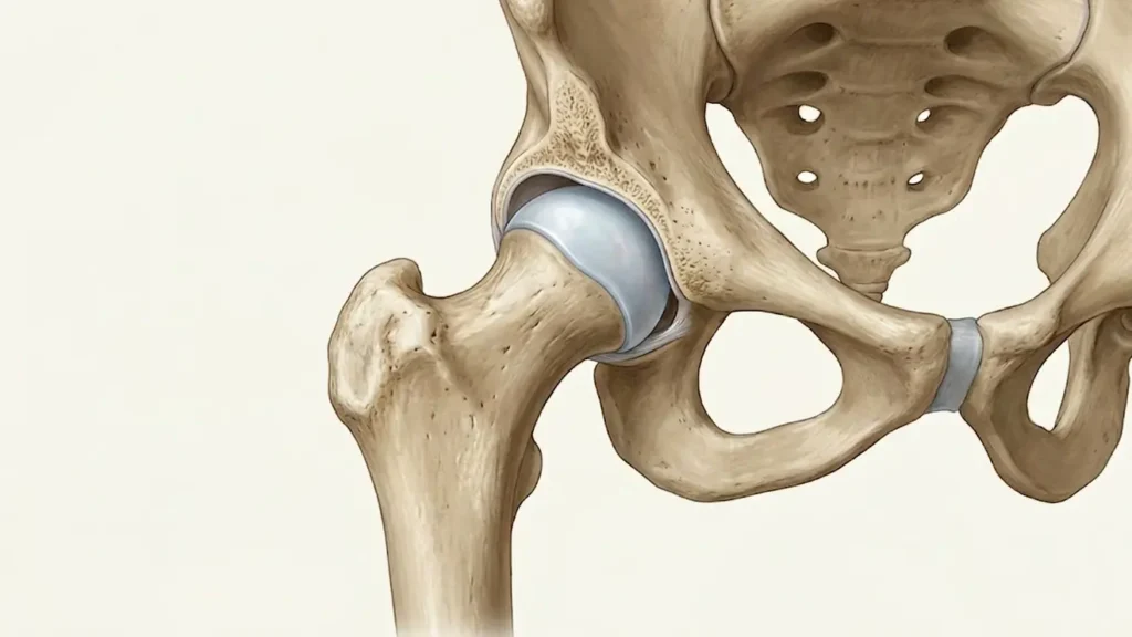
To understand why radiology is so critical, we must first look at the anatomy of a healthy hip. The hip is a “ball-and-socket” joint. The “ball” is the femoral head (the top of the thigh bone), and the “socket” is the acetabulum (part of the pelvic bone).
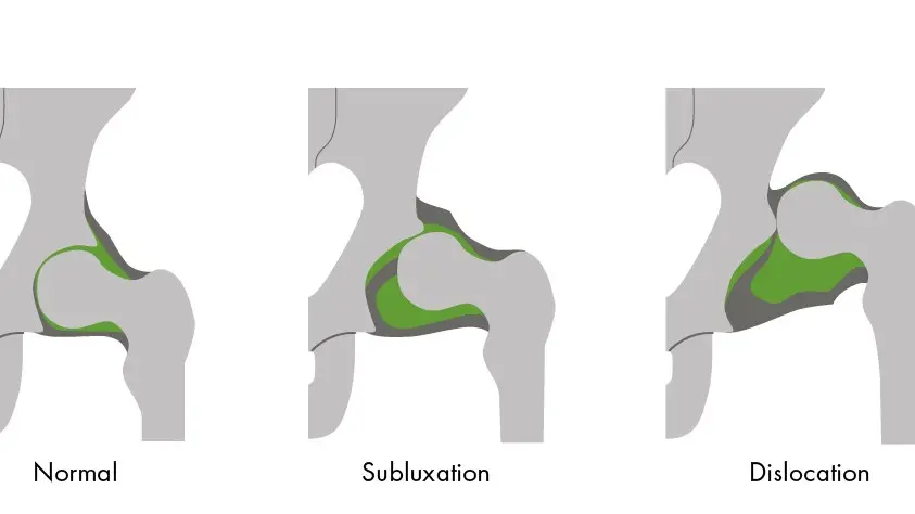
In a perfectly functioning joint, the femoral head fits snugly into the acetabulum, held in place by strong ligaments and a rim of cartilage called the labrum. This alignment allows for a wide range of motion, walking, running, and jumping. However, in cases of congenital hip dislocation, this relationship is disrupted. If the socket is too shallow or the ball is unstable, the joint cannot develop correctly, leading to long-term gait issues and premature arthritis if left unaddressed.
What is a Congenital Dislocation of the Hip?
Congenital dislocation of hip (now more commonly referred to as Developmental Dysplasia of the Hip or DDH) describes a spectrum of conditions where the hip joint does not develop properly. It ranges from a mild “click” in the hip to a complete separation of the joint. In the UAE, where family health is a top priority, routine screenings are standard, yet many cases still require the keen eye of a specialist to interpret subtle radiographic signs.
Diagnosing Congenital Hip Dislocation: Physical Examination
Before a single image is taken, a pediatric specialist will perform a physical exam. In the first few months of life, doctors use the Barlow and Ortolani maneuvers. These tests involve gently moving the infant’s legs to feel if the hip “pops” out of the socket or “clicks” back in.
However, physical exams have limitations. They are highly dependent on the skill of the examiner and the relaxation of the infant. This is where congenital hip dislocation radiology becomes the definitive tool for confirmation.
Radiographic Features: Congenital Hip Dislocation Radiology
Radiology is the backbone of orthopedic diagnosis. Depending on the age of the child, different tools are utilized to visualize the internal structures of the hip. The primary goal of congenital hip dislocation radiology is to assess the “coverage” of the femoral head by the acetabulum and to check for any delay in bone development.
Ultrasound: The Neonatal Standard
For infants under four to six months of age, congenital hip dislocation ultrasound is the gold standard. Because an infant’s hip is mostly made of cartilage (which does not show up well on X-rays), ultrasound uses sound waves to create a clear picture of the soft tissues.
Radiologists look for the “Alpha Angle” a measurement of the depth of the socket. A shallow socket (low alpha angle) indicates hip dysplasia. Ultrasound is non-invasive, radiation-free, and allows the doctor to see the hip move in real-time, providing a “stress test” for the joint.
Plain Radiography: Landmarks and Lines
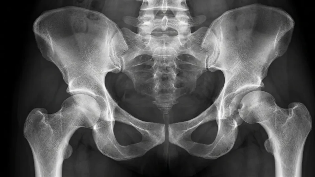
Once a child reaches six months of age, the bones begin to ossify (harden), making hip dislocation radiology via X-ray more effective. When interpreting a congenital dislocation of hip X-ray, specialists look for several key landmarks:
- Hilgenreiner’s Line: A horizontal line drawn through the pelvic cartilage.
- Perkin’s Line: A vertical line perpendicular to Hilgenreiner’s line. In a normal hip, the femoral head should sit in the lower-inner quadrant formed by these lines.
- Shenton’s Line: A smooth arc that should continue from the inner thigh to the pelvis. A “broken” Shenton’s line is a classic sign of dislocation.
Advanced Imaging: CT and MRI
In complex cases, or when a child is already in a spica cast after surgery, advanced imaging is required.
- CT Scans: Often used to confirm that the hip is properly reduced (put back in place) while the child is in a cast.
- MRI: Offers the most detailed view of the labrum, cartilage, and surrounding muscles without the use of radiation. It is particularly useful for identifying “obstacles” to reduction, such as fatty tissue or tight tendons blocking the socket.
How is Congenital Hip Dislocation Treated?
The treatment of hip joint dislocation treatment depends entirely on the age of the child and the severity seen on the scans.
- Pavlik Harness: For newborns, a soft brace called a Pavlik harness is used to hold the hips in a flexed and open position, allowing the socket to deepen naturally.
- Closed Reduction: If the harness fails, a doctor may manually move the hip back into place under anesthesia.
- Open Surgery: In older children or severe cases, surgery is required to reshape the socket or the femur.
Radiology's Role in Management
Radiology is not just for the initial diagnosis; it is the roadmap for the entire treatment journey. Periodic congenital hip dislocation ultrasound or X-ray sessions ensure that the harness is working and that the hip remains stable as the child grows. Without consistent radiographic follow-up, there is a risk of “relapse,” where the hip slowly slips out of place again.
How Can Dr. Usama Saleh Help You?
When it comes to your child’s mobility, there is no room for error. Dr. Usama Saleh’s website is a premier resource for parents in Dubai and Sharjah who are seeking specialized care for pediatric orthopedic conditions. As a leading expert in the UAE, Dr. Usama combines clinical precision with a compassionate approach, ensuring that every family feels supported.
Whether you have been told your child has a “hip click” or you are seeking a second opinion on a hip dislocation radiology report, Dr. Usama Saleh provides the expertise needed to navigate these complex diagnoses. By utilizing the latest imaging technology and evidence-based treatment protocols, he ensures that children receive the best possible start in life.
Book Your Consultation Today
If you are in Dubai or the surrounding areas and have concerns about your child’s hip development, do not wait. Early intervention is the key to avoiding invasive surgeries later in life. Visit Dr. Usama Saleh’s website to learn more about his specialized pediatric services and to schedule a comprehensive evaluation.
Book an Appointment with Dr. Usama Saleh
FAQs about congenital hip dislocation radiology
Conclusion about congenital hip dislocation radiology
Understanding congenital hip dislocation radiology is vital for any parent navigating a DDH diagnosis. From the initial ultrasound that spots a shallow socket to the detailed X-ray landmarks that guide a surgeon’s hand, imaging is the bridge between uncertainty and a successful cure.
In the UAE, families have access to world-class orthopedic care. By choosing a specialist like Dr. Usama Saleh, you are ensuring that your child’s hip health is managed with the highest level of expertise. For more information or to discuss your child’s imaging results, visit Dr. Usama Saleh’s website today. Proper diagnosis today means a more active, pain-free tomorrow for your little one.